A wound in the perineum can follow cancer surgery, severe infection or injury. Reconstruction brings healthy tissue into the area so it can close, stay clean and let you sit, walk and manage hygiene comfortably again.
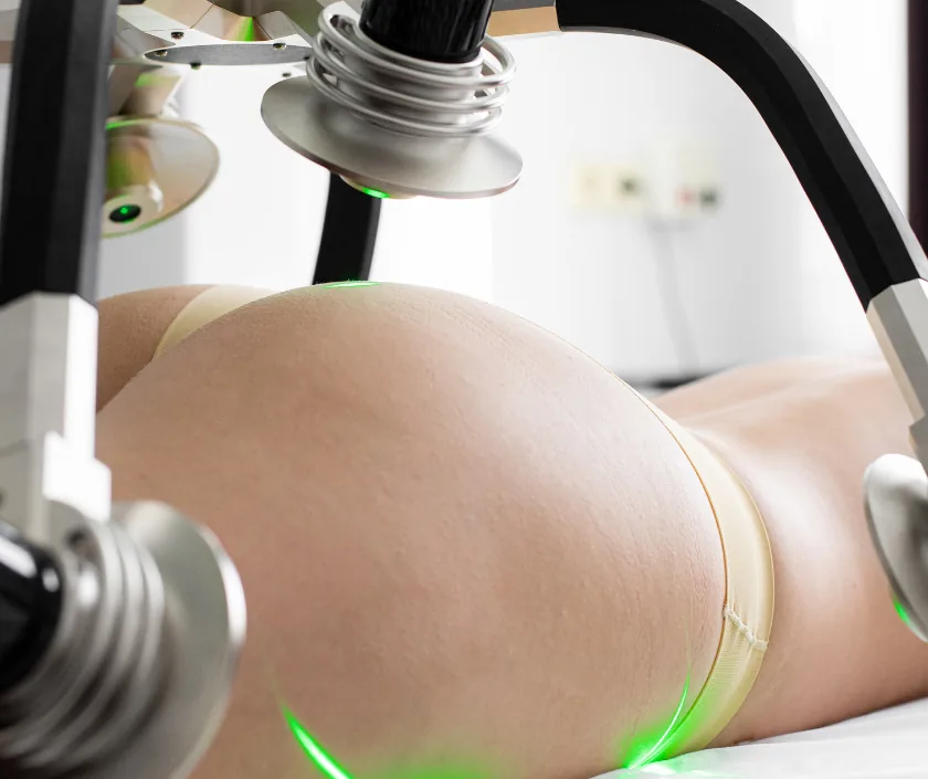
Perineal defect reconstruction closes a wound between the thighs at the base of the pelvis, most often after removal of bowel or genital cancer, or after severe infection. Healthy tissue is brought in from the thigh, buttock or abdomen to fill the space and close the skin. The aim is a clean, healed area that allows sitting and normal hygiene.
The perineum lies between the thighs at the base of the pelvis. It contains the openings of the back passage and the genital tract, so any wound there sits in a warm, moist and mobile place. Those conditions make simple stitching unreliable, particularly after cancer surgery or radiotherapy.
Assessment covers the size and depth of the wound, whether a cavity remains inside the pelvis, and how healthy the surrounding skin is. Previous radiotherapy, a stoma, continence and mobility are all discussed, because each affects the plan and the recovery. Swabs and a scan may be arranged when infection or a deep collection is suspected.
Reconstruction usually relies on a flap. Tissue from the thigh, the buttock or the lower abdomen is moved in with its own blood supply, filling the space inside the pelvis and closing the skin above. Drains are placed, and nursing staff show you how to sit, wash and care for the wound before you leave hospital.
Reconstruction suits people with a perineal wound that will not close on its own, and who are well enough for flap surgery and a careful recovery.
The wound is examined and its depth assessed, with imaging when a cavity remains in the pelvis. Continence, mobility, stoma care and previous radiotherapy are discussed, because each shapes the flap chosen.
Unhealthy tissue, old scar and any infected material are removed until healthy tissue is reached. Samples may be sent for culture so that antibiotics can be targeted properly.
Tissue is raised from the thigh, buttock or lower abdomen with its blood supply preserved. The donor area is chosen to sit away from irradiated skin wherever that is possible.
The flap is moved in to fill the space inside the pelvis and to close the skin without tension. Drains are placed so that fluid cannot gather beneath it.
Sitting, lying and hygiene routines are planned before you leave hospital. Nursing staff show you how to keep the area clean and how to protect the flap as it settles.
Rest and regular position changes protect the flap, with pain relief and drains in place. Nursing staff help with hygiene and show you how to move without dragging on the wound.
Sitting is reintroduced gradually, often with a cushion and for short spells at first. Discharge home follows once the wound is dry and you can manage washing and dressings.
Most people sit and walk far more comfortably. Reviews check the wound and scar, while driving or lighter work is discussed according to progress.
Scars soften and sensation slowly changes. Any remaining difficulty with sitting, hygiene or sexual function is discussed openly at review and managed.
Most people achieve a healed perineum and find hygiene and sitting far easier than before surgery. Scars are visible at both the perineum and the donor site, and the area feels different, often with patches of numbness. Sitting comfort improves gradually rather than immediately. Recovery can vary, particularly after radiotherapy, and some wounds need extra time or a small further procedure.
Wounds in this area carry a higher risk of healing problems than elsewhere on the body, and that is discussed frankly beforehand.
Hygiene and position do much of the work at home, and nursing staff go through both with you before discharge.
Wounds in the perineum sit in a moist, mobile place. Without healthy tissue brought in, many stay open for months despite excellent care.
Sitting is reintroduced gradually with guidance and a cushion. Most people manage short spells early and build up steadily from there.
Flaps taken from outside the treated area bring their own blood supply, which is precisely why they work where local closure fails.
Discharge, odour, difficulty sitting and sexual concerns are ordinary clinical matters, and describing them plainly leads to better treatment.
Perineal work needs privacy, plain speaking and practical support, so consultations here are unhurried and a written estimate is issued before admission.

Cost depends on the size and depth of the defect, the flap chosen, whether cancer surgery is being carried out at the same time and the length of stay. A written estimate is prepared after assessment. It separates surgeon and anaesthesia fees, theatre charges, ward stay and review visits, and the team will help with insurance paperwork where cover applies.
These are the questions that come up most often in consultation. If yours is not here, send it on WhatsApp and the team will reply, usually the same day.
Ask your question →A written estimate follows assessment, since the depth of the defect, the flap chosen, any combined cancer surgery and the length of stay all affect the figure. Every element is itemised before admission.
The area is warm, moist and constantly moving, and it lies close to the back passage. After radiotherapy the blood supply is poorer still. Bringing in healthy tissue is usually what allows closure.
Sitting starts in short spells with a cushion and builds up over the following weeks. Progress is checked at review. Recovery can vary, so guidance is tailored rather than fixed to a set date.
Plain water and gentle patting dry are usually all that is advised, along with loose cotton clothing. Nursing staff go through the routine before discharge and answer questions at each dressing visit.
Some change is possible, and much depends on the original surgery rather than the reconstruction itself. Concerns are discussed openly at review, and practical advice or referral is offered when it helps.
Often yes, and planning it together tends to give the best healing. The cancer team and reconstructive surgeon agree the plan in advance so both operations run in one session.
Bring recent scans, operation notes, biopsy and radiotherapy records, and a list of medicines. Details of stoma care or continence are useful too. A written estimate follows once the plan is agreed.
Seeing patients from across the city and beyond: read about the practice on the plastic surgeon in Surat page, or check what a written estimate covers on the costs and insurance page.
