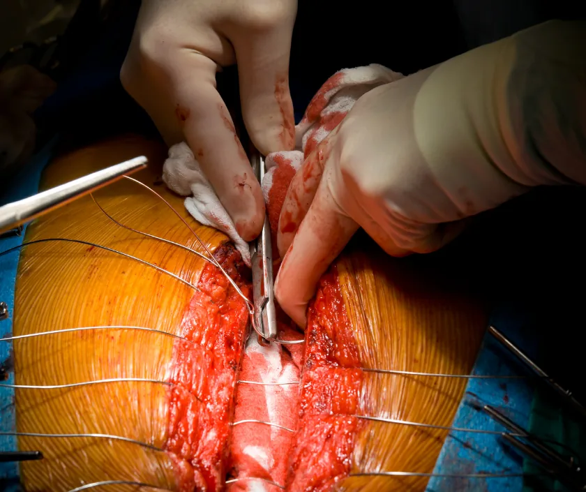
The chest wall is a moving cage of ribs, breastbone and muscle that protects the heart and lungs. When part of it breaks down, the problem is rarely only skin deep. A wound that opens after heart surgery, a rib segment removed with a tumour, or a deep burn can all leave the chest unstable, leaking or exposed to infection.
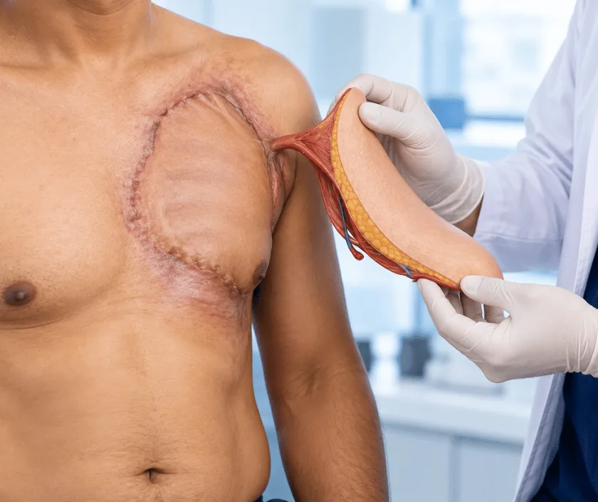
Repair has two separate jobs. The first is to make the cage firm again so that breathing is efficient and the lungs are not sucked inward with each breath. Next comes bringing in healthy, well supplied tissue that fills dead space and seals the area against infection. Muscle from the chest or back, tissue from the abdomen and sometimes a mesh or plate are used together. Planning is shared with the cardiac, thoracic or cancer team so that one operation solves several problems.
Chest wall defects are grouped by their cause and by how much of the bony cage is missing, because those two things decide whether support is needed as well as cover.


The pectoralis major, the latissimus dorsi from the back and the rectus abdominis from the upper abdomen are the usual workhorses. Each can be swung in on its own blood vessels to fill a cavity, and the choice depends on which vessels are still intact.
The apron of fatty tissue inside the abdomen can be brought up to the chest through a small opening in the diaphragm. It moulds into awkward spaces, tolerates infection well and is useful when chest muscles have already been used or damaged.
When several neighbouring ribs are gone, soft cover alone lets the chest move inward on breathing. A synthetic mesh, a mesh and cement sandwich, or titanium rib plates can restore firmness. Rigid material is avoided when infection is still active.
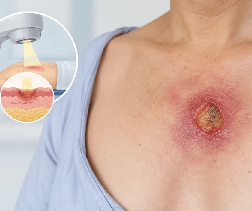
A sealed sponge dressing connected to gentle suction draws the wound edges together, removes fluid and reduces dressing changes. It is a bridge that prepares an infected chest wound for definitive closure rather than a treatment on its own.
Chest wall problems sit close to the heart and lungs, so certain signs should be seen the same day rather than at the next appointment.
Patients referred after heart surgery, injury or cancer treatment usually ask the following.
Ask your question →The estimate depends on how many stages are needed, whether mesh or plates are used, intensive care requirements and hospital stay. Figures are given in writing after assessment. Repairs following heart surgery or cancer treatment are often reimbursable, so carry your insurance file.
It is done routinely in people recovering from cardiac surgery, with the heart team involved throughout. Fitness for anaesthesia, lung function and infection markers are checked first. Risks rise with poor sugar control, smoking and long standing lung disease, and these are addressed beforehand.
Most people spend some days in hospital, longer if the wound was infected or intensive care is needed. Heavy lifting and driving are avoided while the chest knits. Recovery can vary, and breathing exercises are continued at home for several weeks.
Yes, scars are unavoidable, and their position depends on which muscle was moved. They usually fade and flatten over the first year. Where possible incisions are placed so that clothing covers them, and scar care is discussed at follow up.
People with an open, infected or unstable chest wall, and those left with a defect after tumour removal, are usually candidates. Suitability depends on general fitness, lung reserve and whether the underlying disease is controlled rather than on age alone.
Oozing from a breastbone wound should be assessed promptly, because infection can track down to bone and to the space around the heart. Early cleaning is simpler than late reconstruction, so an urgent appointment is safer than watching it at home.
The wound is examined, swabs may be taken and previous operation notes and scans are reviewed. A chest scan is often arranged. The plan, likely number of stages and the recovery path are then explained, along with realistic expectations and risks.
