The outer ear is built early in pregnancy from several small hillocks of tissue that fold and fuse. When that folding goes only part of the way, a baby is born with an ear that is small, tight, buried or set away from the head. Some of these differences also involve the ear canal, so hearing has to be tested even when the outside looks like the only issue.
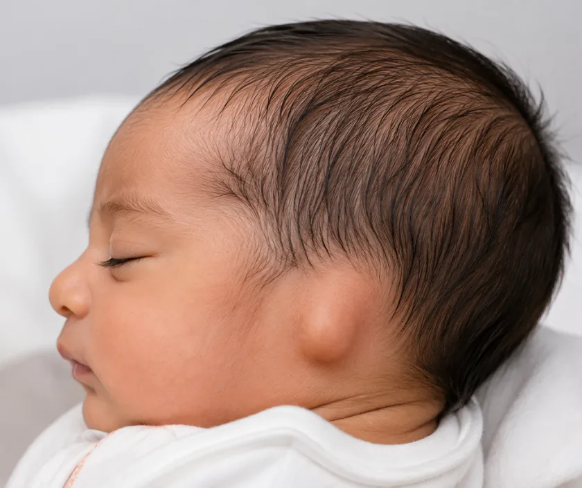
Families usually arrive with two separate worries: will my child hear, and will my child be teased. Both deserve straight answers, and they are handled by different parts of the plan. Hearing is checked in the first months by an ear specialist and an audiologist. Appearance is addressed later, once the ear has grown enough to work with. This page explains the main groups so the words used in clinic make sense, and Elegance Clinic in Surat assesses each child individually.
Ear differences range from a fold that failed to form to an outer ear that never developed. Knowing the group explains why one child is offered moulding and another waits until school age.
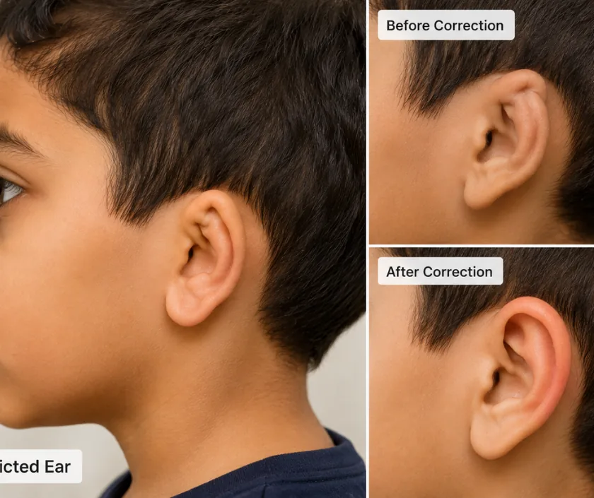
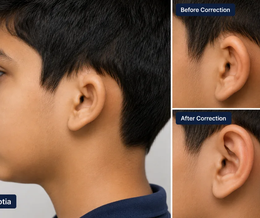
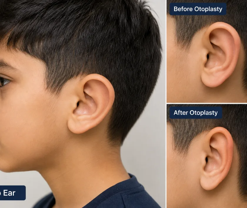
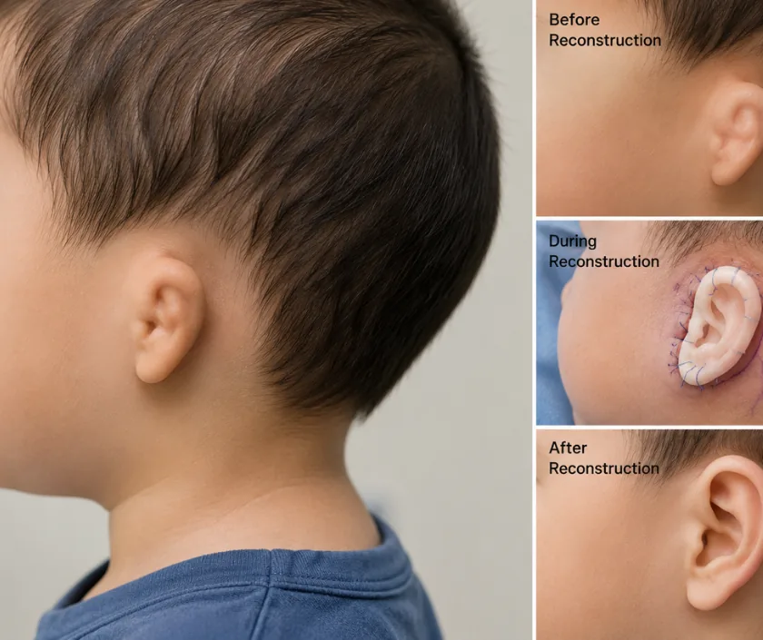
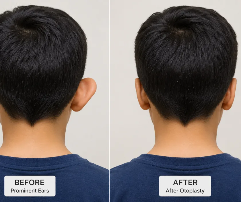
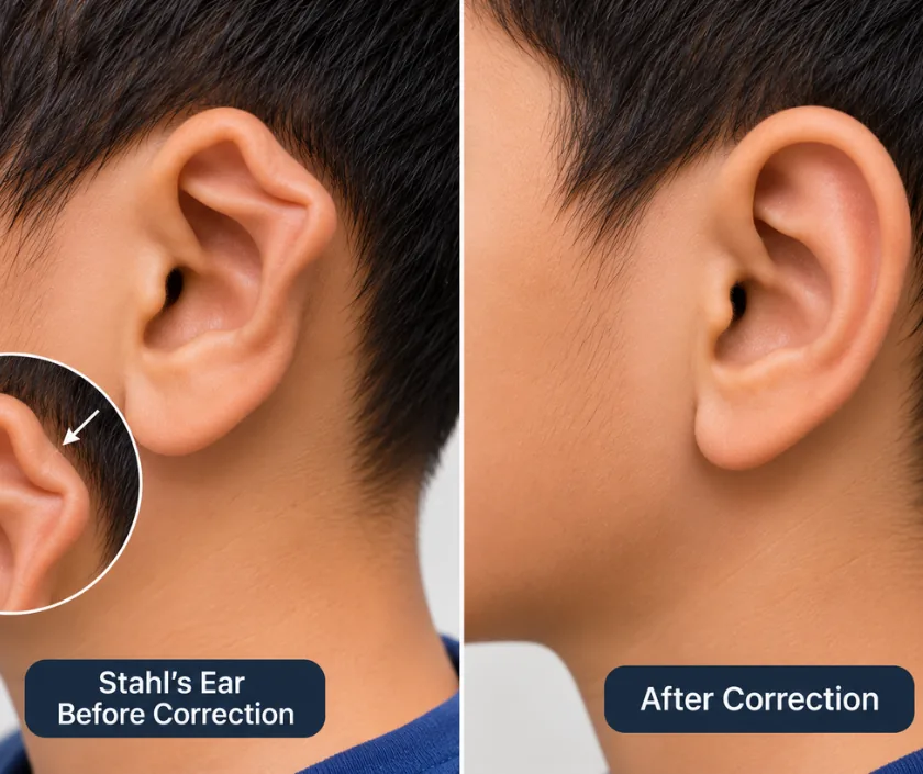
Cartilage stays soft for a short window after birth because of hormones passed from the mother. During those early weeks a gentle splint can reshape a folded rim or a prominent ear without any operation. This window closes quickly, so a newborn ear concern is worth showing to a surgeon promptly.
A narrow or missing canal on one side still allows speech to develop through the other ear, but hearing must be measured rather than assumed. Testing is arranged in the first months. Bone conduction devices are sometimes used so that a child hears clearly during the years when language is forming.
A rebuilt ear needs a supporting frame under the skin. One route carves that frame from the child rib cartilage, which the body accepts as its own. Another uses a porous synthetic frame in a single stage. Each suits different ages and situations, and both are explained fully.
Children start noticing differences at about the age they join school, and comments from classmates often prompt the first visit. Surgery is planned so the ear is grown enough to match the other side, while still landing before those comments settle into something a child carries.
Most ear differences are addressed on a planned timetable. These situations are better reviewed quickly.
Answers to the points families raise most often. An examination is still needed before anything is planned for your child.
Ask your question →Charges vary with the number of stages, the framework material and the hospital stay involved. Rib cartilage work usually spans more than one operation, so the total is spread out. A written estimate is shared after assessment, and insurance sometimes contributes when hearing is affected.
This has been done for many years and the ribs regrow their shape as the child develops. Chest discomfort for a while afterwards is expected and is managed with pain relief. The team explains the small risks, including air leak and scarring, before you agree.
A head bandage stays on for a short spell and is replaced by a soft band worn at night for some weeks. Swelling and tenderness settle steadily. Children generally return to school quickly, while contact sports and swimming wait until the surgeon gives the go ahead.
The aim is a close match in position, size and outline that passes unnoticed under hair and in photographs. Fine detail differs from a natural ear, and the reconstructed side stays firmer to touch. Realistic pictures of earlier work are shown so expectations stay grounded.
Reconstruction waits until the opposite ear is close to adult size and the chest can spare cartilage, which lands around early school years. Prominent ear correction can be considered a little earlier. Moulding is the exception and belongs to the first weeks after birth.
Not always. The inner ear usually develops separately and often works well, so the loss is in getting sound through rather than sensing it. Testing is arranged early because that answer changes the plan. Hearing on the other side is checked with equal care.
Bring any hearing test reports, birth records and previous scans, plus photographs of the ear from early infancy if you have them. Examination takes only a few minutes. The rest of the visit is spent explaining the stages and agreeing a timetable that suits your family.
