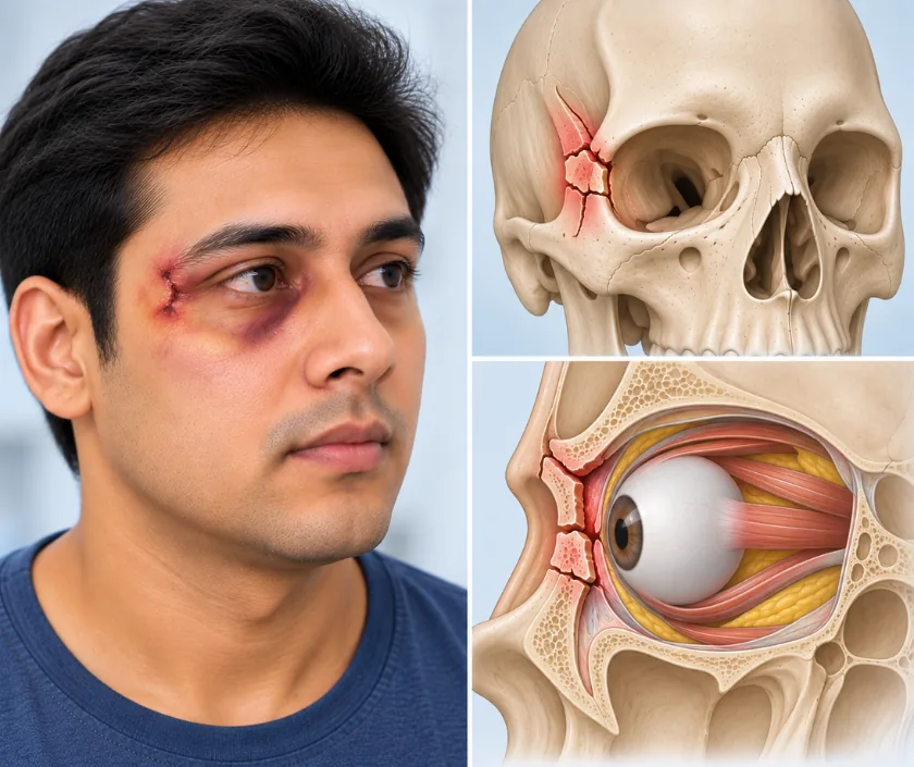
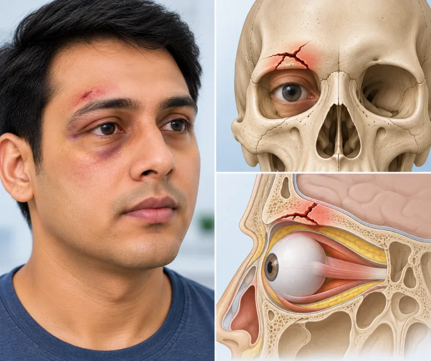
The eye sits inside a bony cone with walls of very different thickness. Its rim in front is strong, while the floor and inner wall are as thin as an eggshell. That design absorbs a blow by letting the thin walls give way, which often protects the eye itself but leaves the socket enlarged. Fat and muscle can then slip into the sinus below, pulling the eye backwards or downwards.
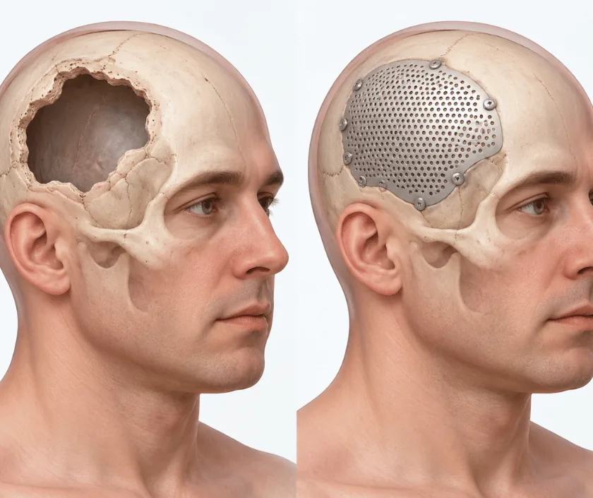
Orbital injuries therefore need two separate questions answered. First, is the eye itself damaged, which an ophthalmologist checks. Second, has the socket changed shape enough to affect vision or eye position. Assessment at Elegance Clinic in Surat combines examination of eye movements, sensation and globe position with a fine cut scan through the sockets. Some injuries settle without surgery. Others are better repaired within the first two weeks, before scar tissue makes the anatomy harder to restore.
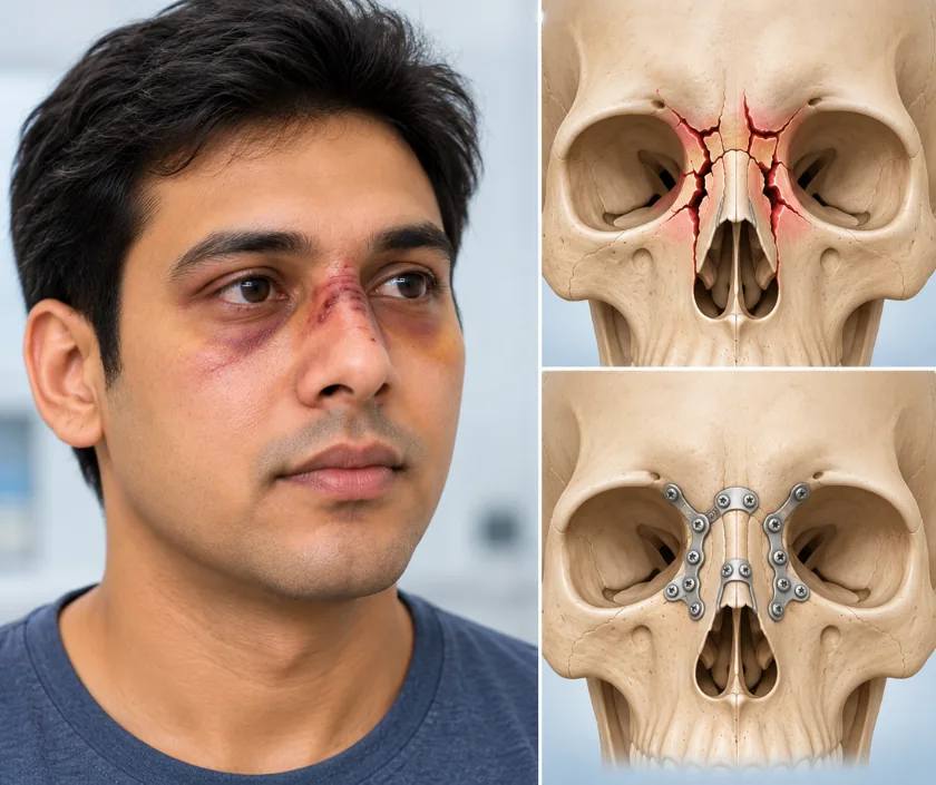
Orbital injuries are grouped by which wall or rim has failed, because that decides the effect on eye position, movement and sensation.
Orthoptists chart eye movements in each direction and measure how far images separate. Repeating that chart over a fortnight shows whether double vision is improving on its own. Those measurements often decide whether an operation is needed at all.
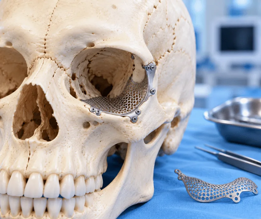
Thin sheets of titanium, porous polymer or resorbable material are used to span a defect, and sometimes the patient's own bone. Choice depends on defect size, whether infection risk is raised and how much support the back of the socket still provides.
The nerve supplying the cheek, side of the nose and upper teeth runs through the orbital floor and is commonly bruised. Numbness or tingling is expected early and usually improves over weeks to months, though some altered sensation can persist.
The socket floor forms the roof of a sinus, so nose blowing can push air into the eyelid tissues. Avoiding forceful blowing for a few weeks is advised, and any decongestant or antibiotic is prescribed rather than started at home.
The eye tolerates little delay. Attend hospital straight away if any of these appear after a facial injury.
← Back to Maxillofacial Trauma & Facial Skeletal Reconstruction
Answers to the questions asked most often after an eye socket fracture.
Ask your question →Charges reflect the implant material, theatre and anaesthetic time, the scans required and whether an ophthalmologist is involved. Custom planned reconstruction for a late deformity costs more than a straightforward floor repair. A written estimate follows assessment.
Working around the eye demands care, and the risks are explained honestly. They include bleeding behind the eye, infection, altered eyelid position, persistent double vision and, very rarely, loss of vision. Vision is checked closely in the hours after surgery.
Eyelid swelling and bruising are marked for one to two weeks, then fade. Double vision often improves across the same period as swelling settles. Heavy lifting, nose blowing and contact sport are avoided for several weeks after the repair.
The aim is symmetry that reads as normal in conversation. A slight difference in eye position or eyelid crease may remain, particularly after severe injury. Photographs before and after help judge the change against realistic expectations.
No. Small fractures with full eye movement, no meaningful double vision and normal eye position are often watched for a couple of weeks and improve on their own. Surgery is advised when tissue is trapped or the eye has clearly sunk.
A trapped muscle, particularly in children, needs release urgently. Most other repairs are done within about two weeks, while the tissues are still mobile. Delay beyond that makes accurate rebuilding of the socket considerably harder.
Vision, eye movement, globe position and facial sensation are tested, and scans are reviewed together with you. An orthoptic assessment may be arranged. The choice between observation and surgery is then explained with the reasoning behind it.
