Marks made of blood vessels are common in newborns, and the words used for them cause endless confusion. Strawberry mark, angioma, port wine stain and birthmark all get applied to very different things. The distinction that actually matters is between a growth of vessel cells, which appears after birth and later shrinks, and a malformation, which is present from the start and stays for life.
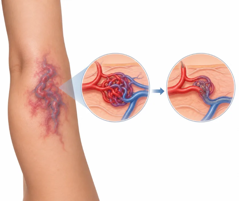
That single split changes everything: whether to watch and wait, whether medicine helps, whether laser or injections are the right route, and whether surgery is needed at all. Getting the label right early spares families years of unnecessary worry, and occasionally it catches a lesion that needs prompt treatment near an eye or an airway. This page explains the standard grouping in plain terms. Elegance Clinic in Surat examines these marks and arranges scans where the diagnosis is not obvious.
Modern classification separates growths of vessel cells from malformed vessels, and then sorts malformations by the type of channel and the speed of flow. Treatment follows directly from that grouping.
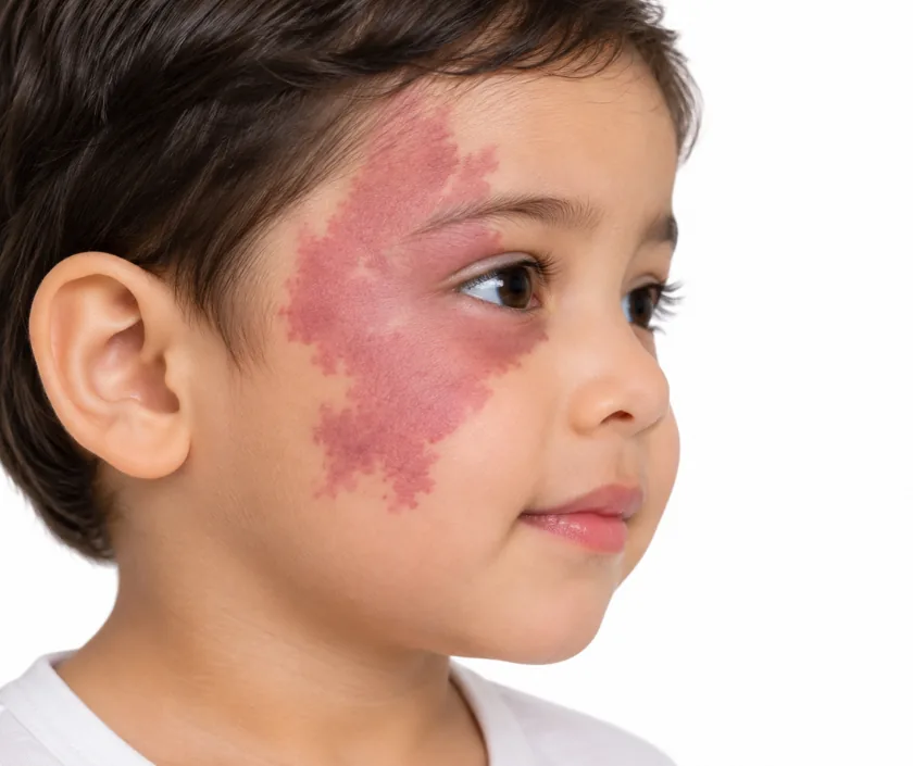
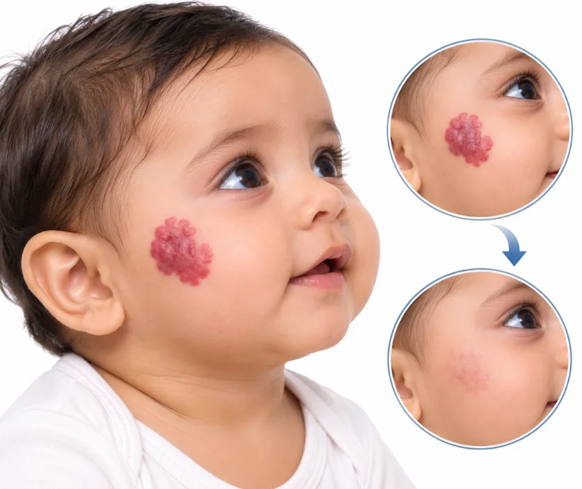
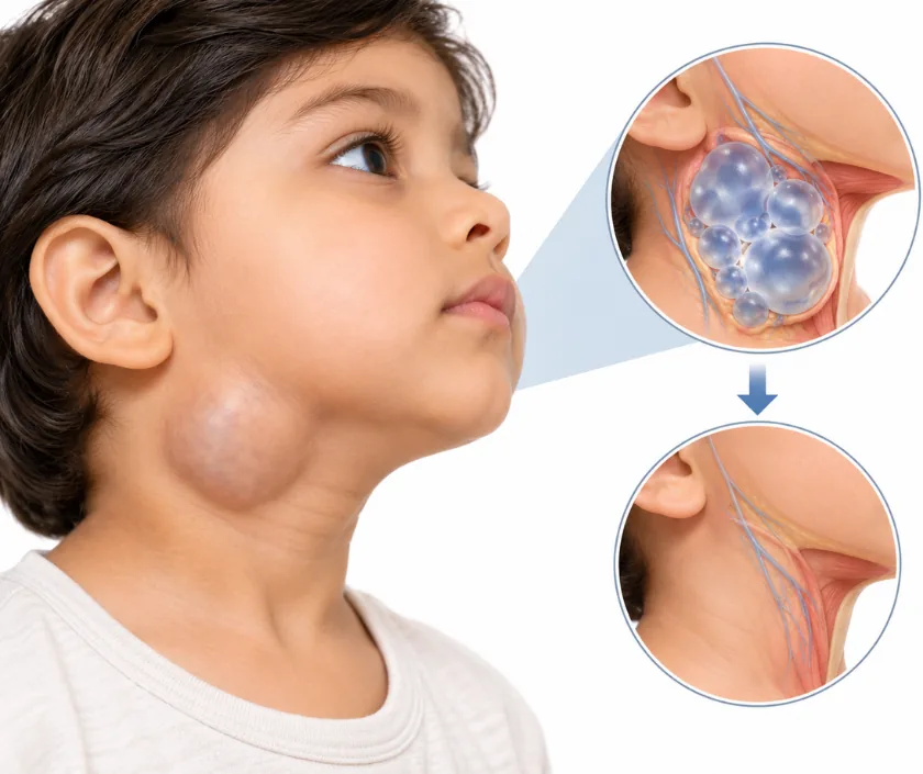
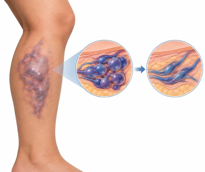
Families often arrive having been told several different words for one mark. A haemangioma that will fade and a malformation that will not can look similar in a photograph taken at a few months. Sorting the two out determines whether the honest advice is patience or treatment.
A great many of these marks are diagnosed by looking and by asking how they behaved over the first weeks. Ultrasound with flow measurement is added when the lesion sits deep or the story is unclear. Detailed imaging is reserved for planning treatment of larger malformations.
Laser targets the colour in blood vessels and works best on flat red marks. Several sessions are spaced across months, with cooling and, for young children, sedation or anaesthesia. Skin looks bruised for a while afterwards. Fading is gradual, and some marks need maintenance sessions later.
A haemangioma in a nappy area or a skin fold can break down and become raw, which is painful and slow to settle. Gentle cleaning, prescribed dressings and prompt review help it heal. Any mark that bleeds repeatedly should be assessed rather than simply covered.
Most vascular marks are harmless and can be reviewed at a routine visit. These situations deserve an earlier appointment.
Answers to the concerns families bring most often. Examination remains the only way to label a particular mark.
Ask your question →It varies widely with the diagnosis. Observation costs nothing beyond review visits, laser is charged per session, and injection treatment or surgery for a malformation carries hospital and anaesthesia charges. An estimate is prepared once the type is confirmed, listing the likely number of sittings.
Vascular laser has been used on infants for many years and settings are adjusted for skin tone and age. Expect bruising for a week or two afterwards. Blistering, temporary lightening or darkening of the skin can occur, and these possibilities are explained before the first session.
Fading after laser builds session by session rather than appearing at once, with several weeks between sittings. Injection treatment for a venous malformation needs swelling to settle before the result shows. Patience matters here, and photographs at each visit make slow progress easier to see.
Many infantile haemangiomas fade a great deal on their own, though loose skin or a faint blush may remain where a large one once sat. Port wine stains lighten substantially with laser rather than vanishing. Realistic aims are agreed at the outset and reviewed as treatment goes on.
Treatment helps at older ages too, although a patch that has thickened over the years usually needs more sessions than one treated young. Response is assessed after the first few sittings. Adults with these marks are treated as well, often for the same reasons.
Urgency arises when a growing lesion blocks vision, interferes with feeding, sits near the airway, bleeds repeatedly or has ulcerated. Any of these needs review within days rather than months. A stable, flat mark on the trunk or limb almost never needs rushing.
The mark is examined and felt for warmth and pulsation, and you will be asked how it looked at birth and how it has changed since. Bring early photographs, since they often settle the diagnosis. An ultrasound may be arranged before any plan is confirmed.
