The facial skeleton is built as a set of struts and buttresses that carry chewing forces up into the skull and protect the eyes, airway and brain. When enough force is applied, those struts break in patterns that repeat from patient to patient. Recognising the pattern tells the surgeon which structures are at risk, what the bite will do, and whether an operation is needed at all.
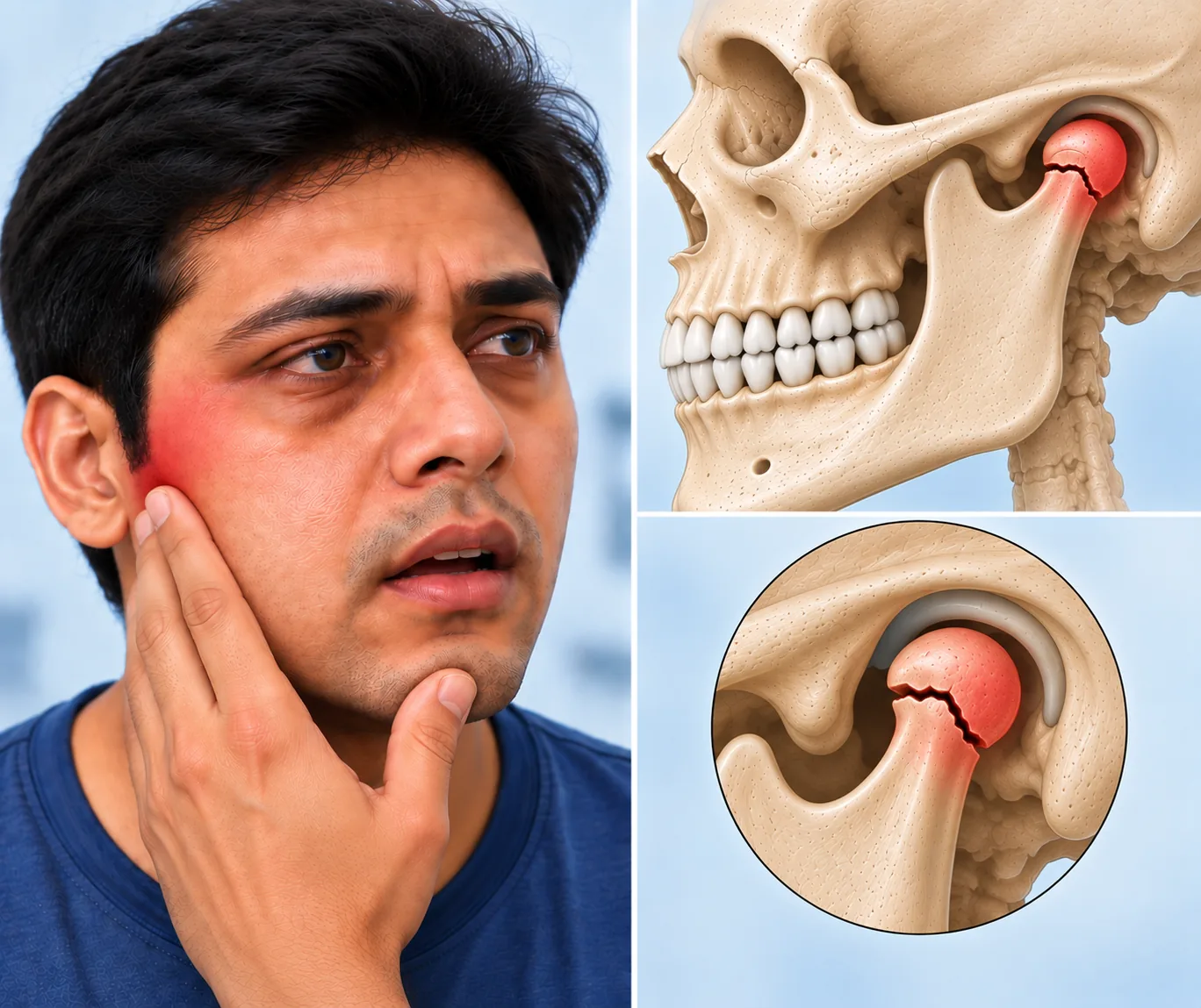
This page sets out the main facial fracture patterns and how they are treated. Diagnosis begins with examination of the bite, eye movements, facial sensation and any step in the bone edges, followed by a scan that shows the fracture in three dimensions. Treatment at Elegance Clinic in Surat ranges from observation and a soft diet to plates and screws placed through hidden incisions. Our Mandible Fracture page covers lower jaw injuries in detail, and the same principles of restoring the bite first apply across the whole face.
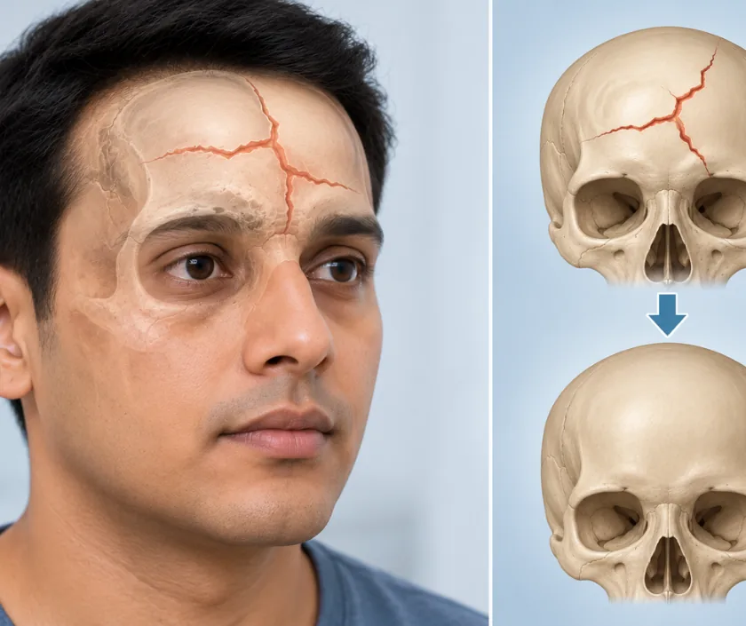
Facial bones break in recognisable patterns. Naming the pattern predicts which functions are affected and how the repair will be approached.
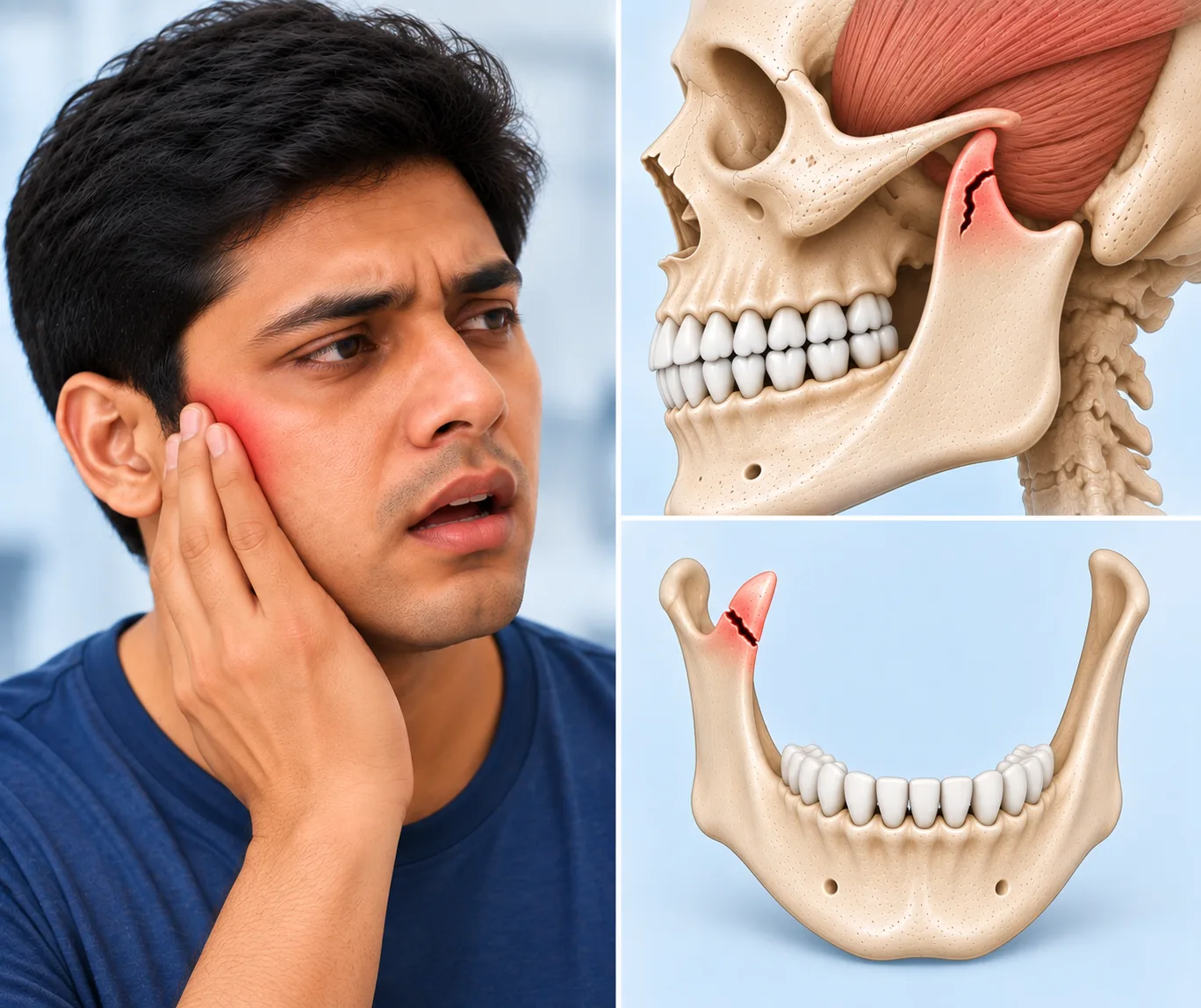
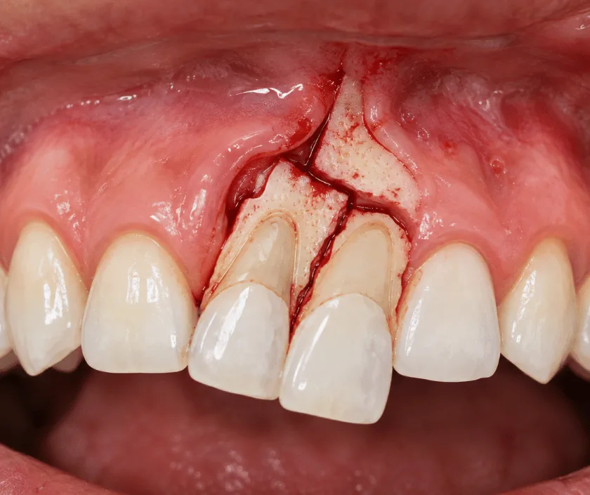
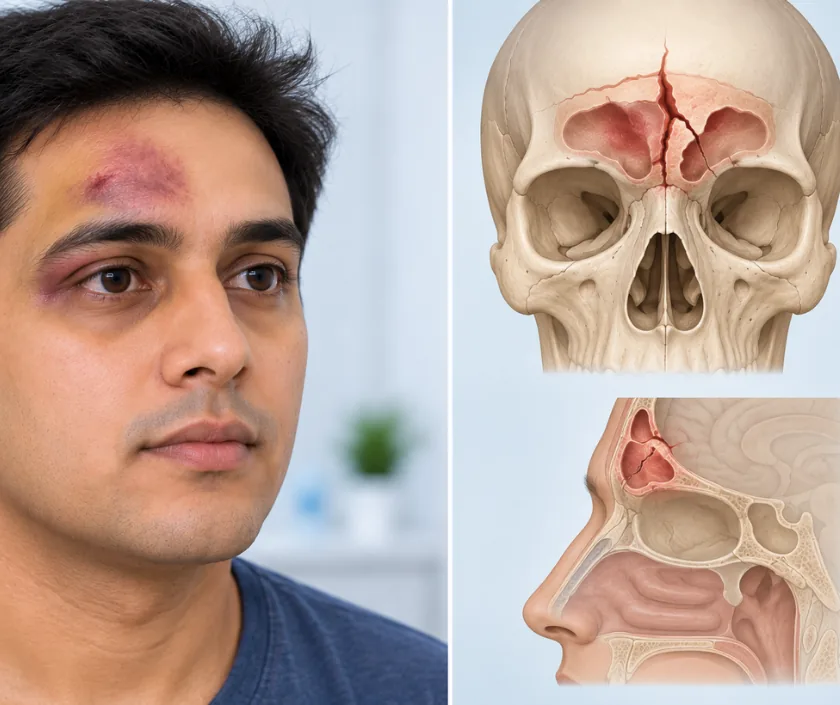
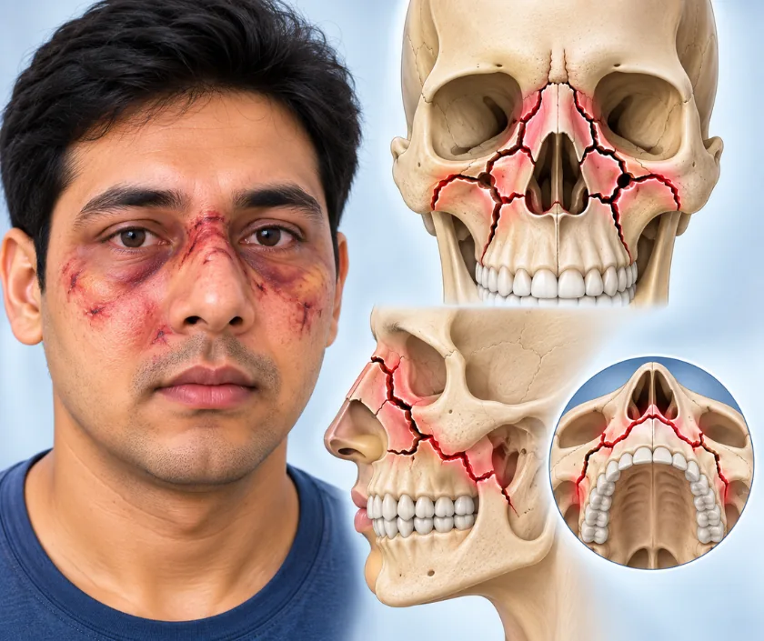

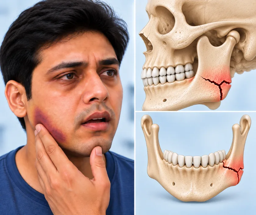
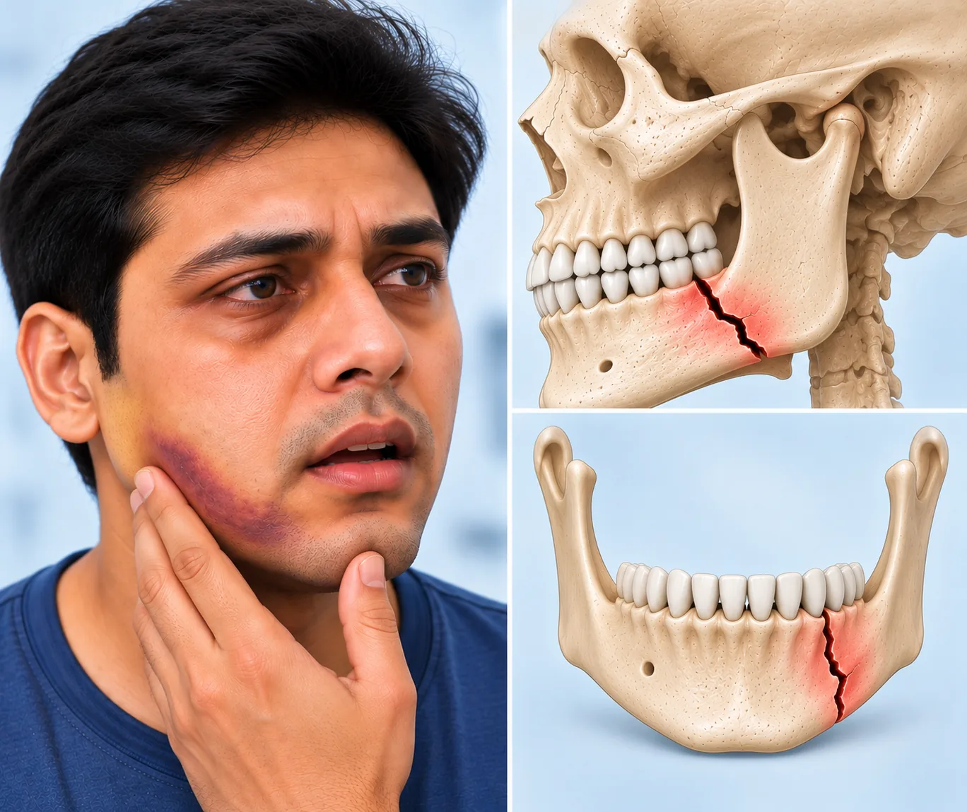
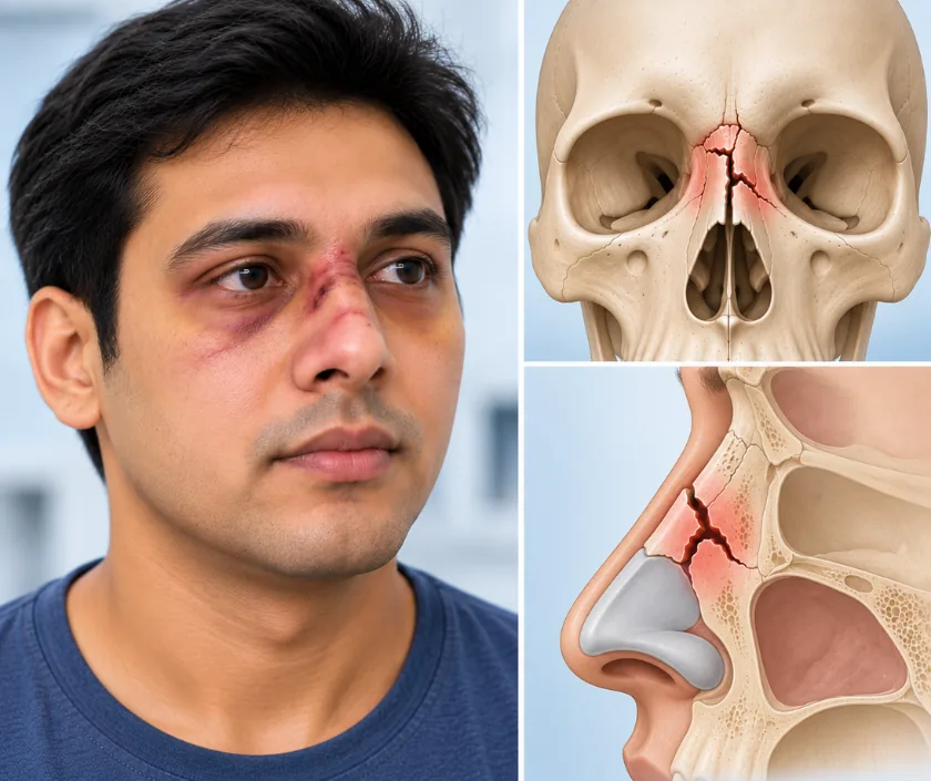
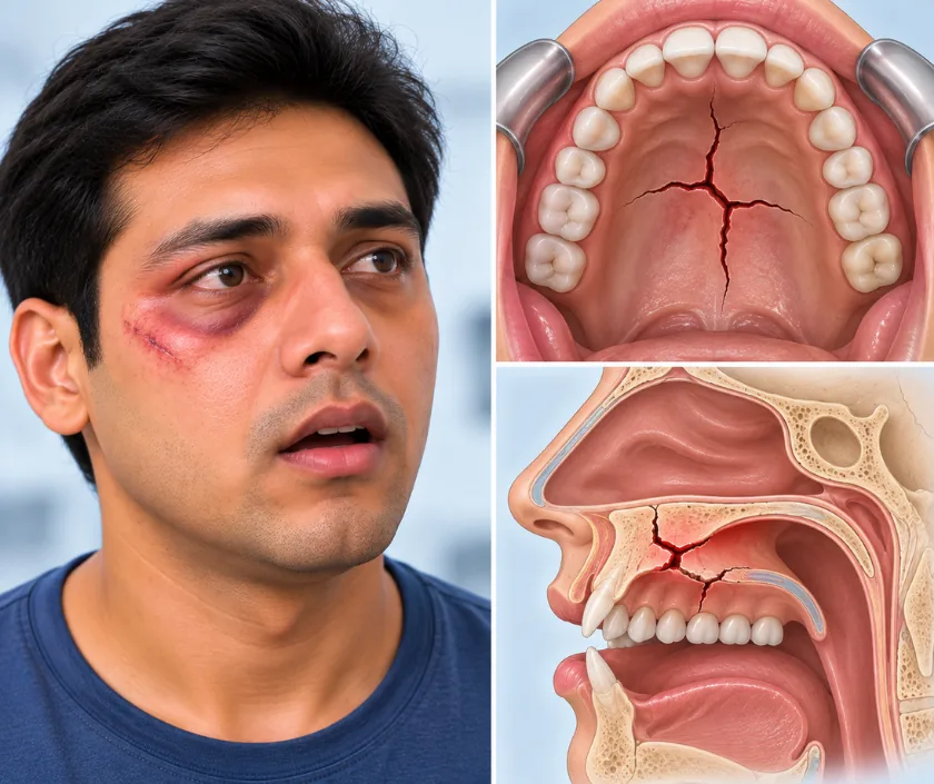
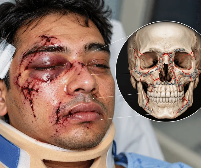
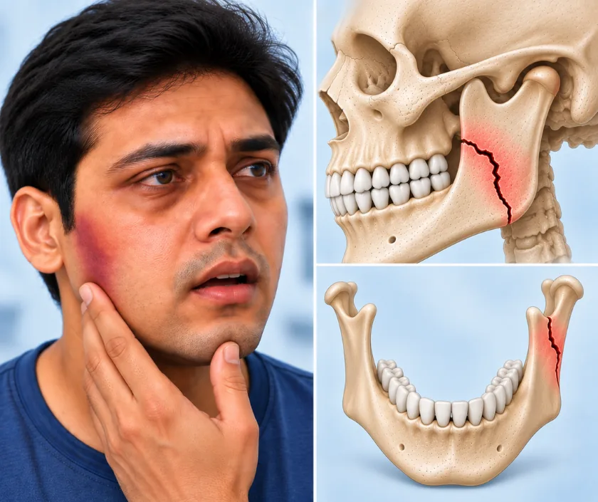
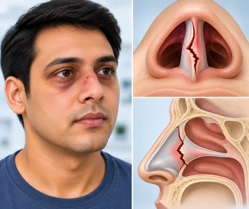
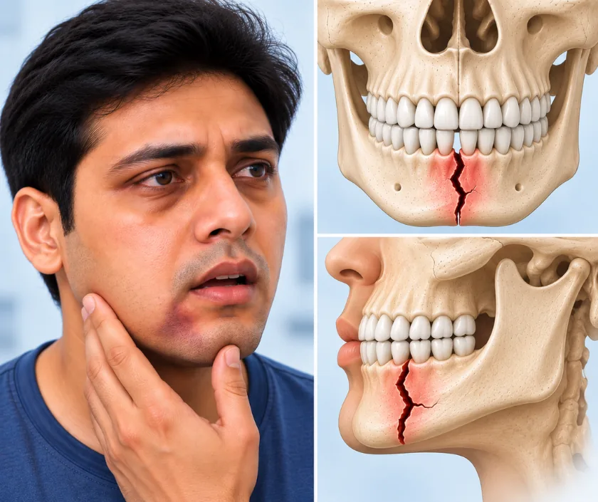
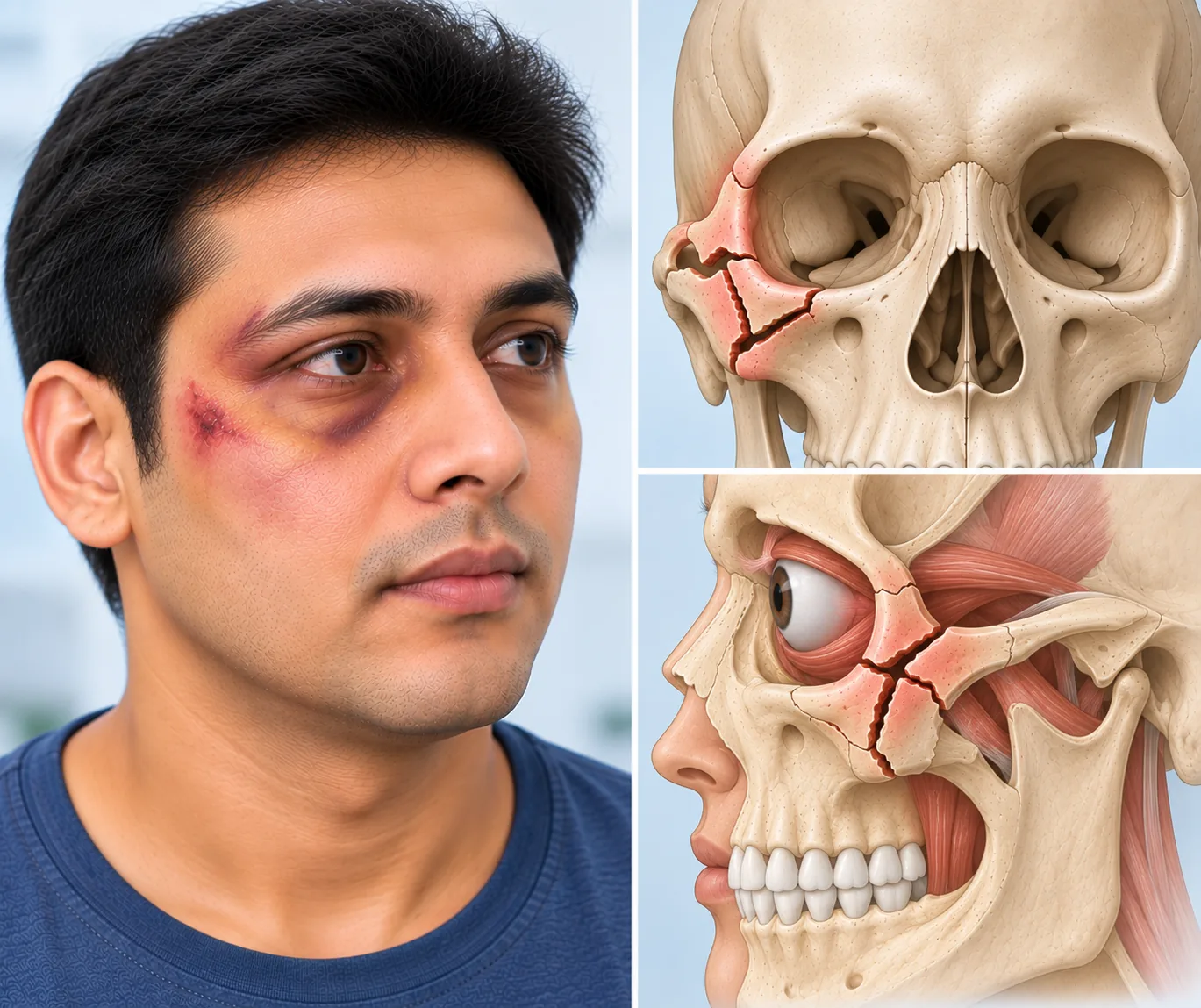
Teeth meet in a pattern unique to each person, and even a small step is felt immediately. Restoring that bite comes before restoring the outline of the face, because bone set in the wrong position leaves chewing uncomfortable however good the profile looks.
Nerves running through the cheekbone and jaw are often bruised or stretched, leaving numbness of the cheek, lip or gum. Sensation usually improves over weeks to months, though recovery can vary. Persistent numbness is reviewed rather than simply accepted.
Bone needs time and steadiness to unite. Soft or liquid diets, avoiding hard foods, and careful mouth hygiene protect the repair. Where fixation is placed between the jaws, specific instructions and equipment are given before you leave hospital.
A fracture that healed in the wrong position can leave a flat cheek, a sunken eye, a crooked nose or an uneven bite. Correction later involves recutting and repositioning the bone, which is more involved than fixing the injury at the time.
Some signs after a facial injury point to a fracture that should not wait.
← Back to Maxillofacial Trauma & Facial Skeletal Reconstruction
Practical answers to what patients ask after a facial injury.
Ask your question →Cost depends on how many bones are involved, the plates and implants used, theatre and anaesthetic time and the length of stay. A single nasal correction sits at one end of the range and a multiple bone repair at the other.
Fixation of facial fractures is routine and well established, with risks including bleeding, infection, plate irritation, nerve numbness and, in orbital work, changes to vision. Every risk is discussed against the consequences of leaving the fracture untreated.
Swelling and bruising dominate the first two weeks. Bone union takes around six weeks, during which diet and activity are restricted. Contact sport is usually avoided for longer. Numbness and final contour can take several months to settle.
Repair aims to restore the pre injury shape and function as closely as possible. Small differences in cheek projection, nasal line or eye position can remain, particularly in severe injuries. Old photographs are useful for planning and for judging the result.
No. Undisplaced fractures with a normal bite, normal eye movement and acceptable contour are often managed with observation, a soft diet and review. Operation is advised when the bite is off, vision is affected or the shape is clearly altered.
Difficulty breathing, heavy bleeding, loss of vision or double vision, clear fluid from the nose or ear, and a jaw that cannot close all need immediate hospital care. Other fractures are usually repaired within the first week or two.
The bite, eye movements, facial sensation and bone edges are examined, and a scan is reviewed or arranged. Findings are shown on the images, options and timing are explained, and old photographs are used to plan the intended result.
