The heel takes the first shock of every step and has very little spare tissue to lose. When an ulcer there reaches the heel pad or the bone beneath it, reconstruction is what keeps the foot worth walking on.
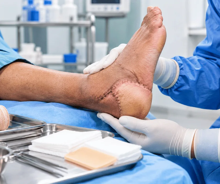
Heel ulcer reconstruction rebuilds the padded cover over the back of the foot after a deep ulcer has destroyed it. Dead tissue and any infected part of the heel bone are removed, then a flap of living tissue is brought in to cover the area, because a graft alone rarely survives in that spot.
Two forces create heel ulcers. Standing and walking press the heel pad against the ground, while lying in bed presses the same area against a mattress. Add numb nerves and reduced circulation, and skin that is already thin gives way. Since barely any soft tissue sits between skin and heel bone, the ulcer reaches bone quickly.
Once bone is exposed, dressings have nothing to grow over. Surgery removes the dead skin, the unhealthy fat and, where needed, the outer layer or a portion of the heel bone. Whatever remains has to be covered with tissue that carries its own blood supply. A local flap taken from the side of the foot or from the calf works for many defects, while a larger loss may need tissue moved from elsewhere in the body and joined to blood vessels under the microscope.
Closure alone is not the goal. A heel has to tolerate load, friction and the edge of a shoe for years, so the cover chosen must be durable, and the way you sit, sleep and walk afterwards is planned as carefully as the operation itself.
This surgery suits a person whose circulation can support a flap and whose heel still has enough bone left to be useful. Careful selection matters more here than anywhere else in the foot.
Pulses are examined and vascular studies are done, since the hindfoot has fewer routes for blood than the front of the foot. Any correctable narrowing is treated before reconstruction is planned.
Dead skin, unhealthy fat and infected bone are cleared under anaesthesia. Samples go for culture so antibiotics can be aimed accurately, and the clean out may be repeated before closure.
Size and depth of the defect guide the choice. A local flap from the foot or calf suits many heels, while extensive loss may need free tissue joined to vessels under the microscope.
Tissue is inset without tension and the heel is shaped so that no ridge or fold presses into a shoe later. Drains and a splint protect the repair through the first days.
Afterwards the heel is kept clear of every surface, including the bed, and weight returns only in stages. Pressure relieving cushions and footwear are arranged before discharge.
The leg is elevated and the flap is checked regularly for colour and warmth. No pressure at all is allowed on the heel, including while you are lying down.
Dressings are changed and the donor area is watched as well. Moving about happens with a walker or wheelchair, while a pillow or boot keeps the heel floating clear.
Most flaps have settled by now. Standing and short walks begin under guidance, often in a heel relief shoe, and the skin is inspected closely for early redness.
Cover toughens and becomes able to take normal load. Moulded footwear, pressure care in bed and regular checks continue to protect the rebuilt heel.
A rebuilt heel is sturdy, though it is not the heel you were born with. New cover feels different, often has little sensation, and needs protective footwear for the long term. Many people stand and walk comfortably once the flap has matured. Healing here takes longer than in other parts of the body, and recovery can vary with circulation, infection and how well pressure is avoided.
The hindfoot is a demanding place to rebuild, so the risks are set out plainly before you agree to surgery.
Pressure is the enemy of a healing heel, and most pressure happens quietly at home or in bed.
Once the wound reaches bone it will not close by itself, and surgery is what provides fresh cover.
This area takes load and friction, so a thin graft usually breaks down. Flap tissue is normally required.
Infected bone can often be removed and the heel resurfaced. What matters is how much bone remains behind.
Nerve damage hides deterioration, so a heel ulcer can grow deeper while feeling like almost nothing.
Elegance Clinic in Surat offers flap based heel reconstruction with the vascular assessment that should always come first. Dr. Ashutosh Shah discusses the realistic options, including the times when surgery is not the safer choice.

The cost of heel reconstruction reflects the method of cover, the length of stay and whether bone had to be removed. A local flap is less involved than free tissue transfer, which needs microsurgery and closer monitoring afterwards. A written estimate follows assessment, and insurance cover is checked and explained before admission.
These are the questions that come up most often in consultation. If yours is not here, send it on WhatsApp and the team will reply, usually the same day.
Ask your question →The estimate depends on the type of flap, the length of hospital stay and whether infected bone must be removed. A written figure is shared after examination and vascular tests. Insurance cover is checked in advance, so there are no surprises at discharge.
Any flap can fail, and the back of the foot has a less generous blood supply than the front. Circulation is therefore studied before surgery is offered. Risks, alternatives and the option of not operating are all explained before you decide.
Weight stays off the heel for several weeks while the flap settles, then returns in stages. Many people walk in a protective shoe by around six weeks, although recovery can vary with circulation, infection and how strictly pressure is avoided.
The rebuilt area usually has little or no sensation, so it feels different from the skin around it. It does become sturdy enough for walking as it matures. Daily visual checks then replace the warning that feeling would normally give.
Shallow ulcers can heal with offloading, dressings and good sugar control. Once tendon or bone lies exposed, dressings alone rarely succeed. Surgery is then offered because solid cover protects the bone underneath from infection.
Ask for review as soon as a heel wound appears, changes colour, smells, or fails to shrink over a fortnight. Sudden swelling, warmth or fever needs attention the same day. Early treatment usually means a smaller operation.
Your wound is measured and photographed, pulses and sensation are tested, and imaging or vascular studies are arranged. Swabs guide the choice of antibiotic. Findings, reconstructive options and a written estimate are then discussed with you.
Each technique below has its own page explaining how it works and when it is chosen.
Seeing patients from across the city and beyond: read about the practice on the plastic surgeon in Surat page, or check what a written estimate covers on the costs and insurance page.
