Under the ball of every foot sit five rounded bone ends that share the load of walking. When one of them drops or becomes prominent, it presses through the skin and creates an ulcer that keeps coming back.
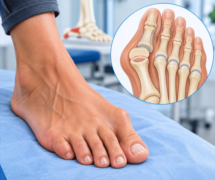
Metatarsal head resection removes the prominent end of a forefoot bone that is pressing through the sole. Taking away that bump removes the point of concentrated pressure, so the ulcer beneath it can heal. The same operation is used when infection has spread into that bone from the wound above.
The forefoot is built to share weight across five bone ends. Clawing of the toes, a tight calf or an earlier operation can drop one of them lower than its neighbours. That single point then meets the ground first and hardest. Skin responds by thickening into callus, the callus presses inward, and an ulcer forms underneath it.
Dressings treat the surface while the bone keeps pressing. Casts and insoles do help by spreading the load, although only while they are being worn. When an ulcer under one bone end refuses to close, or when infection has entered that bone, removing the prominent head becomes the logical step.
The operation itself is straightforward. Through a small incision on the top of the foot, or through the wound, the rounded end of the bone is removed and the area is washed out. Bone goes for testing when infection is suspected. Sometimes more than one head is removed to keep the forefoot balanced, since leaving a prominent neighbour simply moves the problem along.
It suits a person whose forefoot ulcer sits over one identifiable bone end, with enough circulation to heal a wound in that part of the foot.
Examination, callus pattern and imaging pinpoint which bone end is causing the pressure. Whether one head or several need attention is decided at this stage.
Where infection is spreading, cleaning and antibiotics come first. When infection sits inside the bone itself, resection becomes part of the treatment rather than a delay to it.
Through a small incision, the rounded end of the bone is removed and the space is washed thoroughly. Samples go to the laboratory whenever infection is suspected.
Neighbouring bone ends are assessed, and occasionally more than one is trimmed, so the remaining bones share load evenly instead of creating a fresh peak.
The ulcer is then dressed or closed. A protective shoe or cast keeps weight away while skin closes over the space left behind.
Elevation controls swelling and simple pain relief is usually enough. Walking is limited to short distances in the protective shoe provided.
The wound is checked and the ulcer measured. Most ulcers start to shrink noticeably once the bone beneath them has gone.
Healing is often complete, and the protective shoe gives way to fitted footwear with a moulded insole that spreads load across the forefoot.
The foot settles into its new pressure pattern. Reviews watch the neighbouring bone ends, since they now carry a share of what was removed.
Ulcers under the resected bone usually heal well once the pressure has gone, and many people walk comfortably in fitted shoes afterwards. The forefoot is slightly narrower at that point and push off feels a little different. Because load has to go somewhere, a new ulcer can develop under a neighbouring bone end. Insoles and regular checks are what keep that risk low.
Moving pressure is the whole purpose here, and it brings possibilities of its own that deserve a plain explanation.
The bone is gone, yet the walking habits that created the ulcer are still there and need managing.
Most people walk normally afterwards, since the remaining bones share the load between them.
Dressings cannot overcome bone pressing outward. Taking the pressure away is what allows closure.
Load moves to the neighbours, so footwear and regular checks continue in order to protect them.
Nerve damage removes the pain entirely, and an ulcer that feels like nothing may still be sitting on infected bone.
Elegance Clinic in Surat identifies the exact bone causing the pressure before operating, and considers the whole forefoot rather than one point. Dr. Ashutosh Shah plans the resection so the remaining bones share load evenly.

Because this is a short procedure with a brief stay, the estimate is usually modest. What moves the figure is how many bone ends are treated, whether infected bone is being cleared and how the ulcer above is closed. A written estimate follows assessment, and insurance cover is confirmed before admission.
These are the questions that come up most often in consultation. If yours is not here, send it on WhatsApp and the team will reply, usually the same day.
Ask your question →The estimate is usually modest, since the procedure is short and the stay is brief. It rises when several bone ends are treated or infected bone must be cleared. A written estimate is given after assessment, and insurance cover is confirmed in advance.
The forefoot copes well when one bone end is removed, because the remaining bones take up the load. Circulation and infection are checked first, and the plan aims to leave a balanced forefoot rather than a new pressure peak.
Walking usually starts within a few days in a protective shoe, with the wound settling over the following weeks. Fitted footwear follows once healing is complete. Recovery can vary with the size of the ulcer and how well circulation supports it.
That is the main long term risk, because load must transfer to the neighbouring bones. Balancing the forefoot during surgery and wearing moulded insoles afterwards reduce the chance considerably, and reviews catch early changes.
Offloading, dressings and callus care are always tried first, and sometimes a calf lengthening resolves the pressure without touching bone. Resection is considered once those measures fail, or when the bone underneath is already infected.
Raise it when an ulcer has not closed with proper offloading over several weeks, keeps returning, or begins to discharge. Acting before infection settles into bone usually keeps the operation small and the recovery straightforward.
The sole is examined for callus and prominence, the ulcer is measured and probed, and pulses and sensation are tested. Imaging shows which bone is involved. Options and a written estimate are then explained to you and your family.
Seeing patients from across the city and beyond: read about the practice on the plastic surgeon in Surat page, or check what a written estimate covers on the costs and insurance page.
