In diabetes, an infection of the skin and deeper soft tissue can travel a long way before it causes much pain. Early drainage, careful antibiotics and firm sugar control together decide how much of the foot is saved.
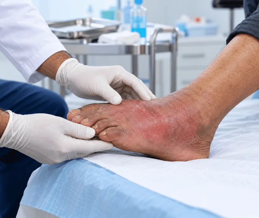
Diabetic soft tissue infection is infection of the skin, fat, fascia or tendon in someone with diabetes, most often starting from a small foot wound. Reduced sensation hides the pain, and high sugar levels weaken the body's defence, so infection spreads along tissue planes. Treatment combines urgent drainage of pus, removal of dead tissue, targeted antibiotics, tight sugar control and protection of the foot from pressure.
Assessment looks past the small opening on the surface. The foot is examined for swelling, tenderness along the tendons, discharge and loss of sensation, and the pulses are checked. Blood tests, a sugar profile and an X ray or scan help show whether infection has reached bone or a deep space. A probe that reaches bone through the wound is a significant finding.
Surgery follows quickly when pus is present. Deep spaces are opened and drained, dead tissue and infected tendon are removed, and deep samples are sent for culture. Wounds are left open so they drain freely, and a second look in theatre is often planned.
Medical care runs alongside and matters just as much. Sugar levels are brought under control with insulin, antibiotics are narrowed once cultures return, and anaemia and low protein are corrected. Once the wound is clean, cover is achieved with dressings, a skin graft or a flap, and protective footwear is fitted before discharge.
Drainage and debridement suit anyone with a deep or spreading diabetic infection. What follows depends on blood supply, the extent of tissue loss and how well sugar can be controlled.
The wound is probed, the foot examined for deep tenderness, and pulses and sensation are tested. Blood tests, sugar profile and imaging are arranged, and admission is advised when swelling extends beyond the wound.
Deep spaces are opened, pus is released and dead skin, fat, fascia and tendon are removed until healthy tissue is reached. Deep samples go for culture, and the wound is left open.
Insulin is used to steady blood sugar, antibiotics are adjusted once culture results arrive, and anaemia, kidney function and nutrition are corrected. A physician reviews the person daily during admission.
Regular dressings, or negative pressure therapy for larger wounds, encourage clean granulation tissue. Repeat debridement is done if slough or dead tendon appears at a later dressing.
The wound is closed by direct suture, a skin graft or a local flap once ready. Protective footwear or a cast is fitted, and a foot care routine is taught before discharge.
Swelling and pain settle as the pus drains. Intravenous antibiotics and insulin continue, the foot stays elevated, and dressings are changed daily.
Most people go home with a dressing plan and protective footwear. The wound looks cleaner, and short supervised walking with aids usually begins.
Many wounds are closed or grafted by this point. Standing and walking distance improve, though special footwear is still needed at all times.
Skin toughens and gait steadies. Regular foot checks, sugar control and yearly review continue, since a new wound elsewhere on the foot is always possible.
Outcome depends heavily on how early treatment began and on how well sugar is controlled afterwards. Many feet keep useful walking function, though the shape changes where tissue has been removed and special footwear becomes part of daily life. Sensation seldom returns in a nerve damaged foot, so daily inspection stays necessary. New ulcers can appear at fresh pressure points, and regular review is what catches them early.
Surgery in an infected diabetic foot carries risks that are explained before consent is taken.
Daily habits at home protect the result far more than any single treatment in hospital.
Nerve damage removes the warning of pain, so serious deep infection often causes very little discomfort.
Drugs cannot reach pus under pressure or dead tissue, so drainage and debridement remain the core of treatment.
Pressure crushes healing tissue with every step, which is why special footwear or a cast is prescribed.
The risk factors that caused the first wound remain, so daily inspection and proper footwear continue for life.
Elegance Clinic in Surat manages diabetic soft tissue infection as a joint problem of infection, circulation and sugar control. The plan and its stages are explained to the family in plain language.

Cost is shaped by the depth of infection, the number of theatre visits, the length of admission, antibiotic requirements and the way the wound is finally covered. Dressings, cultures and imaging are itemised separately. A written estimate is prepared once the first assessment and any scan are complete, and it is revised openly if further debridement becomes necessary. Diabetic foot admissions are commonly covered by insurance and government schemes, and the team helps with the approval paperwork.
These are the questions that come up most often in consultation. If yours is not here, send it on WhatsApp and the team will reply, usually the same day.
Ask your question →Nerve damage removes the pain that would normally prompt early treatment, high sugar weakens the body's defence, and reduced blood flow limits healing. Together these allow bacteria to spread deep along tendons before the person notices anything wrong.
Only for early, superficial infection without a collection. Once pus has formed or tissue has died, drugs cannot reach the area, and drainage with removal of dead tissue becomes the treatment that actually controls the infection.
Cost depends on how many procedures are needed, the length of stay, antibiotics and the final wound cover. A written estimate is shared after assessment, and the family is informed before any additional procedure is added to the plan.
Sometimes a toe or a small part of the forefoot must be removed to control infection, particularly when bone is involved. The aim is always to take the least tissue that allows healing while keeping the foot useful for walking.
Healing can vary a great deal. Small wounds may close in a few weeks, while deeper defects need dressings for longer before grafting. Sugar control and blood supply influence the timeline more than the size of the wound.
Age and kidney disease raise the risk, yet leaving infection untreated is more dangerous. Physician and anaesthetic colleagues optimise the person first, and procedures are staged so that each one stays as short as possible.
Expect wound probing, examination of pulses and sensation, blood tests, a sugar profile and often an X ray. The likely number of stages, the plan for taking pressure off the foot and a written estimate are then discussed.
Each technique below has its own page explaining how it works and when it is chosen.
Seeing patients from across the city and beyond: read about the practice on the plastic surgeon in Surat page, or check what a written estimate covers on the costs and insurance page.
