Squamous cell carcinoma behaves more assertively than the basal cell type. It can grow into deeper tissue and, in some cases, reach nearby lymph nodes, so removal with a wider margin is followed by a planned repair.
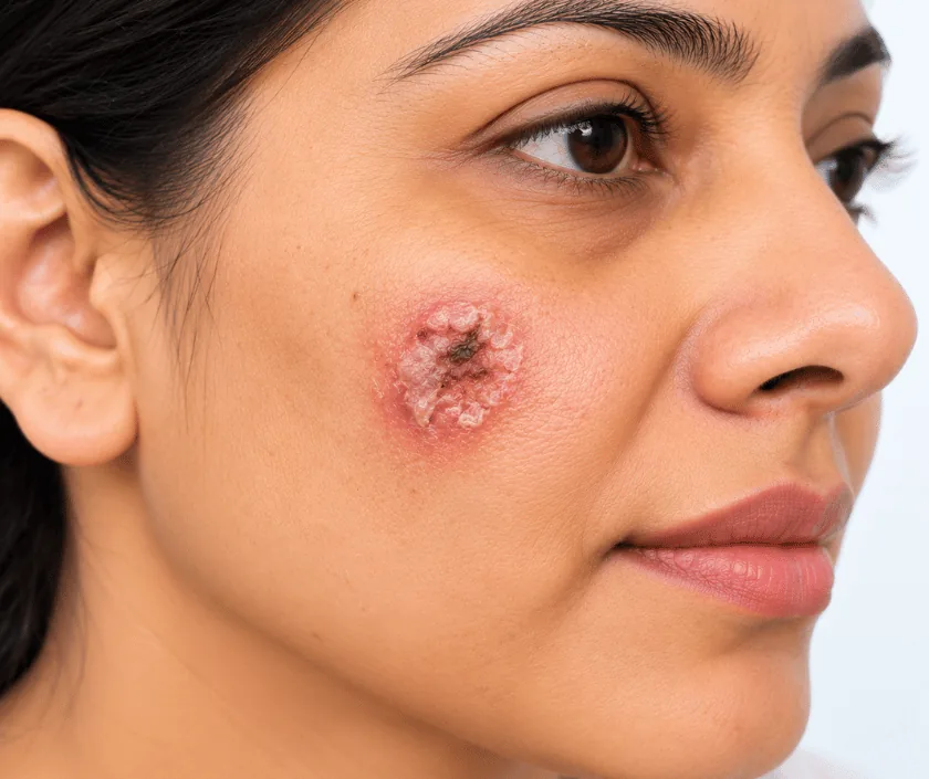
Squamous cell carcinoma reconstruction rebuilds the area after this skin cancer is removed with a wider margin than a basal cell lesion needs. Because it can travel to lymph nodes, the neck, armpit or groin may be examined or scanned. Once margins are confirmed, the gap is closed with a graft or a flap.
Squamous cell carcinoma arises from the flat cells that make up the outer layer of skin. It often shows as a firm, crusted lump or a tender ulcer that grows over weeks rather than years. Sun damaged skin, old burn scars, long standing wounds and areas treated with radiation in the past are common sites.
Because this cancer pushes into deeper layers and can travel through lymph channels, surgery is more thorough than for a basal cell lesion. A wider band of healthy skin is taken, and the depth of removal reaches below the level of the tumour. Lymph nodes in the neck, armpit or groin are examined, and a scan is added when the lesion is large, recurrent or sitting on the lip or ear.
Repair follows once removal is complete. Small gaps close directly, while larger ones need a graft or a flap that brings in skin with its own blood supply. Sometimes the wound is dressed and the repair is done a few days later, after the report confirms clear margins.
Most people with a confirmed squamous cell carcinoma are suitable, and the plan is shaped by how deep the lesion goes and whether nodes are involved.
Biopsy findings are reviewed alongside an examination of the lymph nodes. An ultrasound or scan is added when the lesion is thick, recurrent or sitting in a demanding area such as the lip or ear.
A measured band of healthy skin is marked around the visible edge, wider than a basal cell lesion would need. Depth is planned as well, since this cancer travels downward as much as outward.
Skin, fat and sometimes deeper tissue are lifted as a single specimen with orientation stitches in place. Nodes are sampled when examination or imaging suggested involvement.
Small wounds are closed at once. Larger defects may be dressed for a few days while the report is awaited, so that any repair is built on tissue known to be clear.
Grafts and local flaps rebuild the area, matching thickness and colour as closely as possible. Function comes first around the lip, eyelid and nose, where movement matters more than appearance.
Swelling and soreness peak early, while a dressing protects the graft or flap. Resting the treated part is encouraged, especially on the lower leg.
Stitches are removed and the healed surface is inspected. Pathology is discussed at this visit, along with whether further treatment is needed.
Wounds have usually closed and normal activity resumes. Scars remain pink, and any planned radiotherapy generally starts around this stage.
Colour settles and the repair blends better. Regular skin and node checks continue, because recurrence is most often picked up within the first two years.
Most people heal with a repaired area that works well and looks acceptable, though borrowed skin differs a little in colour and texture. Where the lip, eyelid or nose is involved, some tightness or a slight change in shape can persist. Recovery can vary, and the outcome depends on how deep the lesion was and whether radiotherapy follows.
Risks rise with the size and depth of the lesion, so they are discussed against your own findings rather than in general terms.
The repair needs protection while it takes, and a few simple rules cover most of that.
They can look similar to the eye, though this one grows faster and may reach lymph nodes, which is why margins are wider and nodes are checked.
Depth often surprises people. What shows as a crust can extend well below, so removal is planned in three dimensions rather than two.
Immediate repair suits many cases, yet waiting for the margin report before rebuilding is the safer route when the edges look uncertain.
Review visits watch both the scar and the nearby nodes, since most recurrences appear within the first couple of years.
Elegance Clinic in Surat plans skin cancer removal and reconstruction with the treating oncologist, so margins, node assessment and any radiotherapy fit into a single sequence.

The figure depends on the size and depth of the lesion, whether nodes are sampled, the type of repair and the number of laboratory reports needed. Day care removal with direct closure sits at the lower end, while a flap under general anaesthesia with node surgery sits higher. A written estimate is prepared after assessment.
These are the questions that come up most often in consultation. If yours is not here, send it on WhatsApp and the team will reply, usually the same day.
Ask your question →An estimate is written after assessment, because depth, size, node surgery and the type of repair each change the total. Laboratory reporting is billed separately. Many health policies cover cancer surgery, so bring the policy papers to the first visit.
Yes, it can. Spread happens through lymph channels to nodes in the neck, armpit or groin, more often with thick lesions and with those on the lip or ear. That is exactly why nodes are examined and sometimes scanned.
Growth here is faster and reaches deeper, and small extensions can sit beyond the visible edge. Taking a wider and deeper band lowers the chance of cells being left behind, which reduces the risk of it returning.
Surface healing usually takes about two weeks, and a graft needs protection throughout that period. Most people return to routine work within one to two weeks. Colour and softness keep settling for several months afterwards.
Sometimes. The pathology report describes depth, nerve involvement and margin status, and the oncologist uses those details to advise. When radiotherapy is planned, it usually begins once the wound has healed enough to tolerate it.
Neither is better in general. Grafts suit flat areas with a healthy bed underneath, while flaps bring their own blood supply and suit the nose, lip and eyelid. Site, depth and any planned radiotherapy guide the choice.
Bring the biopsy report and slides, any scans, a list of medicines and details of earlier skin treatments. Photographs showing how the lesion looked when it started are surprisingly useful, so include them if you have any.
Each technique below has its own page explaining how it works and when it is chosen.
Seeing patients from across the city and beyond: read about the practice on the plastic surgeon in Surat page, or check what a written estimate covers on the costs and insurance page.
