Melanoma is the skin cancer that most needs prompt, thorough treatment. Surgery removes a wide band of tissue around the lesion, staging checks whether it has travelled, and reconstruction restores skin cover once the margins are known to be clear.
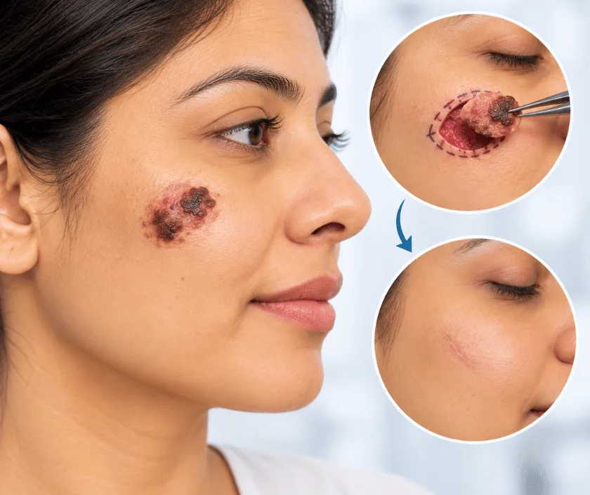
Melanoma excision removes the lesion with a wide margin, and the width is chosen from how thick the melanoma measures under the microscope. Staging often includes a sentinel lymph node biopsy and scans, because melanoma can travel early. Reconstruction with a graft or a flap follows once the margins are confirmed clear.
Melanoma starts in the pigment producing cells of the skin. Many appear as a mole that changes shape, colour or size, while others begin as a new dark spot that looks different from everything around it. Thickness measured on the biopsy matters more than width, because it predicts how likely the disease is to have travelled.
Surgery therefore comes in two parts. First, the area is removed again with a measured wide margin around the original biopsy scar, taken down to the layer beneath the fat. Second, the lymph system is assessed. A dye and a tracer identify the sentinel node, which is taken out through a small separate cut and sent for detailed examination.
Reconstruction follows once everything is known. On the trunk, direct closure often works after the skin is freed. On limbs, face and scalp, a graft or a local flap is usually needed. Where the report shows features that call for more treatment, the medical oncologist joins the plan before the next step is taken.
Surgery suits nearly everyone with a confirmed melanoma, and the real questions are how wide the margin should be and whether the sentinel node needs checking.
Thickness, ulceration and other features on the report decide how wide the margin should be. That same report guides whether a sentinel node biopsy and staging scans are advised, so nothing is chosen by guesswork.
Lymph nodes are examined and scanned where needed. Shortly before surgery, a tracer is injected around the scar so that the sentinel node can be located accurately during the operation.
The old biopsy scar is removed within a measured band of healthy skin, down to the deep layer. Everything comes out as one specimen, marked so the laboratory can orient it correctly.
Through a small separate cut, the first draining node is found with a probe and dye, then removed. Detailed examination of that node shapes the advice you receive afterwards.
The gap closes directly where skin allows, or with a graft or local flap. Comfort and function guide the choice, particularly on the face, the sole of the foot and near joints.
Discomfort is manageable with regular medicine, and dressings stay in place. Movement of the treated limb is kept gentle, so that the repair is protected while it settles.
Stitches come out and the wound is inspected. Reports on the margins and the sentinel node are usually ready, and next steps are agreed at this visit.
Wounds have healed for most people and normal activity returns. Additional treatment advised by the oncologist generally begins around this point.
Scars soften and fade slowly. Skin and node checks continue on a schedule, and you are taught how to examine your own skin between visits.
Wide excision leaves a longer scar than people expect, because the margin is measured from the edge of the original lesion rather than from the biopsy scar. Grafted skin often looks paler or shinier than the skin around it. Recovery can vary with the site, and the outcome depends heavily on what the pathology and node reports show.
The wider the excision and the more the lymph system is disturbed, the more there is to discuss beforehand.
Care at home focuses on protecting the repair and on watching your skin properly.
Taking out a melanoma does not spread it. Delay is the real problem, because thickness increases with time and that measurement drives the outlook.
It is less common in darker skin, yet it does occur, often on the sole, the palm or under a nail, where it can be missed for months.
A diagnostic biopsy is deliberately small. Wide excision afterwards takes the measured margin that the thickness calls for, which is a separate step.
A clear node is reassuring, though it does not end the need for review. Skin checks and node examinations carry on to an agreed schedule.
Melanoma care at Elegance Clinic in Surat is organised with the medical oncologist from the first visit, so excision, node assessment and any further therapy follow one agreed sequence.

Cost is shaped by the width of the excision, whether a sentinel node biopsy is done, the tracer and scans involved and the type of reconstruction. Pathology for melanoma is detailed, so laboratory charges run higher than for other skin lesions. A written estimate is given after assessment and before admission.
These are the questions that come up most often in consultation. If yours is not here, send it on WhatsApp and the team will reply, usually the same day.
Ask your question →A written estimate follows assessment. Margin width, sentinel node biopsy, the tracer, scans and the type of repair all affect it, and melanoma pathology costs more than routine skin reporting. Cover for cancer surgery varies, so bring your policy papers.
It is treated promptly rather than at leisure. Thickness increases with time, and that measurement drives both the margin and the outlook, so wide excision is usually arranged within weeks of the diagnosis.
Margin is measured outward from the edge of the original lesion, not from the biopsy scar, and the skin then has to close in a line. A wider circle of removal therefore becomes a longer straight scar.
It checks the first node that drains the area. Finding cells there changes the advice about further treatment and follow up, while a clear node offers useful reassurance about the stage of the disease.
Sometimes. Thickness, ulceration and node findings guide the medical oncologist, who may advise additional therapy. That discussion happens after the reports arrive, so nothing is decided before the facts are known.
Desk work often resumes within one to two weeks. Physical jobs, gym training and swimming usually wait until around six weeks, particularly after node surgery or a graft on a limb.
Reviews are frequent during the first two years, then spread out. Each visit examines the scar, the skin elsewhere and the lymph node areas, and you are taught how to check yourself between appointments.
Each technique below has its own page explaining how it works and when it is chosen.
Seeing patients from across the city and beyond: read about the practice on the plastic surgeon in Surat page, or check what a written estimate covers on the costs and insurance page.
