Skin treated with radiotherapy years ago can thin, break down and form an ulcer that refuses to heal. Bringing in healthy tissue from outside the treated area is usually what allows the chest to close again.
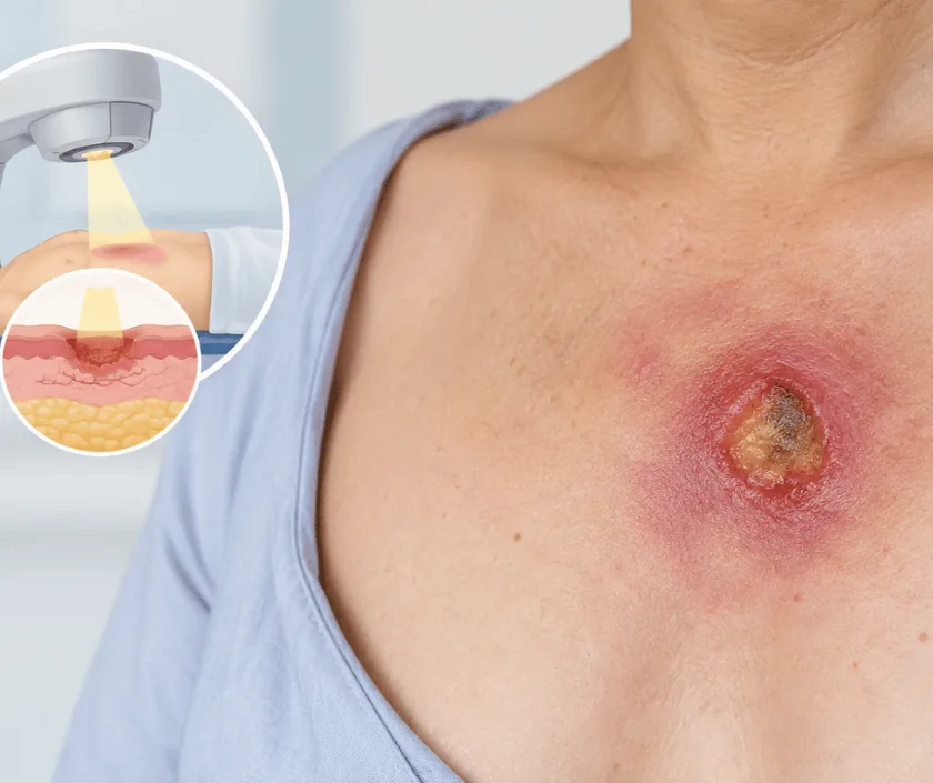
A radiation ulcer on the chest is a wound in skin damaged by earlier radiotherapy. Because blood supply in that area is poor, dressings alone rarely close it. Treatment removes the damaged tissue and covers the area with a flap taken from outside the irradiated field, which brings fresh circulation into a place that had lost it.
Radiotherapy saves lives, yet treated skin pays a price for it. Over months and years the tiny blood vessels in the area narrow, the tissue becomes thin and stiff, and its ability to repair itself falls away. A small knock, a stitch or simple pressure can then open a wound that stays open.
Assessment looks at the whole picture. Your surgeon checks how deep the ulcer runs, whether ribs or an implant are exposed, and how far the radiation damage extends around it. Because a long standing ulcer can occasionally harbour cancer, a biopsy is taken. Swabs, blood tests and a scan may follow when infection or bone involvement is suspected.
Treatment removes the ulcer along with the worst of the surrounding damaged skin. Cover then comes from healthy tissue outside the treated field, usually a muscle or skin flap from the back, the abdomen or the opposite side of the chest. Grafts are avoided over irradiated ground, since they need a healthy bed that is simply not there.
Reconstruction suits people whose chest ulcer has not healed despite careful wound care, and who are well enough for flap surgery with a period of recovery.
The ulcer is examined and measured, and a biopsy is taken from the edge. Imaging may be arranged to check the ribs and deeper tissue. Findings guide whether reconstruction or cancer treatment comes first.
Nutrition, blood sugars and tobacco use are addressed beforehand. Dressings keep the wound clean meanwhile, and any infection is treated so that the operation starts from a better position.
In theatre the ulcer and the worst of the surrounding radiation damaged skin are excised. Generous removal matters, because leaving damaged tissue at the edges usually leads to the wound opening again.
A flap of muscle or skin is raised from outside the treated field and moved across with its blood supply intact. Careful positioning avoids tension, which fragile skin tolerates badly.
Drains stay while fluid settles, and the flap is checked regularly for colour and warmth. Dressings are kept light so that circulation into the new tissue is never squeezed.
You rest with the flap monitored closely. Pain relief, hydration and gentle changes of position protect the new tissue, while drains manage any fluid gathering underneath.
Most people go home once the flap looks settled. Dressing appointments continue, and you are shown how to keep the area clean and free from pressure.
Healing is usually well advanced, though irradiated edges can take longer. Reviews check the wound, and light activity resumes as comfort allows.
Scars mature slowly and colour differences fade only partly. Treated skin nearby still needs protection from sun, friction and minor injury for the long term.
Flap cover usually closes the wound and settles the symptoms that made daily life difficult. The area will look different, with scars at both the chest and the donor site, and colour will not match exactly. Irradiated skin nearby stays fragile, so care is needed with sun, pressure and knocks. Recovery can vary, and some wounds need longer or a second procedure before they close fully.
Surgery in irradiated tissue carries higher risk than surgery in normal skin, and that is discussed frankly before you decide.
Fragile skin needs protecting long after the wound itself has closed.
Changes in the skin can appear years later, because the small blood vessels keep narrowing long after the course has ended.
Dressings help keep a wound clean, yet they cannot supply the blood flow that irradiated tissue has lost.
Grafts need a healthy bed with good circulation. Over irradiated ground they usually fail, so a flap is chosen instead.
Cancer can develop within long standing radiation damage, which is exactly why a biopsy is taken before treatment begins.
Wounds in irradiated skin need patience and planning, so assessment here is thorough, biopsy comes early, and the plan with its written estimate is explained before admission.

Cost depends on the size of the ulcer, the flap chosen for cover, whether a biopsy or further imaging is needed and the length of stay. A written estimate is prepared after assessment rather than quoted in advance. The estimate separates surgeon and anaesthesia fees, theatre charges, hospital stay and follow up visits, and the team can assist with insurance paperwork.
These are the questions that come up most often in consultation. If yours is not here, send it on WhatsApp and the team will reply, usually the same day.
Ask your question →A written estimate follows assessment, since the price depends on the size of the ulcer, the flap needed, biopsy and imaging, and the length of hospital stay. Everything is itemised for you before admission.
Radiotherapy narrows the small blood vessels in treated skin, so the area cannot supply the oxygen and nutrients that repair needs. Dressings keep the wound clean but cannot replace lost circulation.
A biopsy is usually taken from a wound that has been open a long time, because cancer can arise within old radiation damage. Knowing the result early avoids surprises and shapes the treatment plan properly.
Colour and texture rarely match exactly, since tissue is moved from another part of the body. The priority is durable cover over fragile ground. Appearance often improves gradually as scars mature.
Yes, and the plan is coordinated with your oncology team. Where active disease is present, cancer treatment may need to come first. Bring recent scans and reports to the consultation.
Irradiated tissue heals more slowly than normal skin, so healing is judged at review rather than by a fixed date. Many people feel comfortable within weeks, while complete maturing of scars takes longer.
Avoid pressure from straps and tight clothing, keep the skin covered in strong sun, moisturise as advised and report any new breakdown early. Stopping tobacco makes a real difference to healing.
Each technique below has its own page explaining how it works and when it is chosen.
Seeing patients from across the city and beyond: read about the practice on the plastic surgeon in Surat page, or check what a written estimate covers on the costs and insurance page.
