When bone lies open in a wound, the surface has nothing left for skin to grow across. Dressings can keep the area clean, yet closure needs living tissue with its own blood supply moved over the bone.
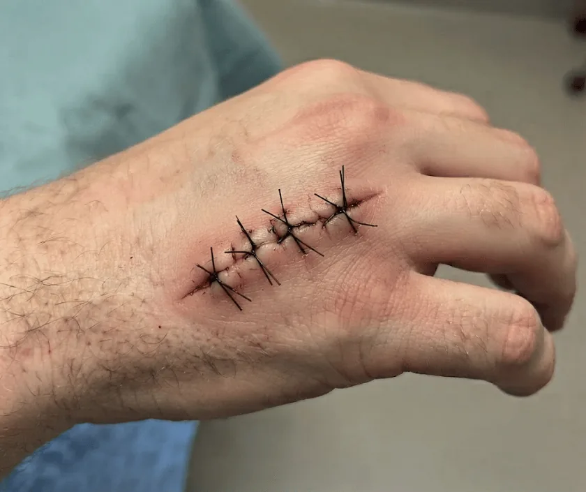
Bone has no skin of its own and no surface that new tissue can creep across. Once it is exposed in a wound, dressings alone will not close the gap, however long they continue. Treatment means removing any dead or infected bone and then covering the area with living tissue moved from nearby or from elsewhere in the body.
Skin heals by growing inwards from the edges of a wound and upwards from the base. Both routes need a soft, moist surface with a blood supply. The outer layer of bone offers neither. It is hard, it has very little surface circulation once its covering has been stripped away, and it dries when left open to air.
So a wound with visible bone behaves unlike other wounds. It may stay the same size for months while dressings change around it. Worse, exposed bone that dries slowly dies, and dead bone becomes a home for bacteria that antibiotics reach poorly. That is how a small open area over the shin or the heel turns into a much larger problem.
The answer is to bring in tissue that carries its own blood supply. Depending on the site and the size, that may be a local flap rotated from beside the wound, a muscle flap, or a free flap taken from another part of the body and joined to vessels under a microscope. First, though, any dead or infected bone has to go, because covering it simply hides the trouble.
A flap is a real operation and it asks something of the body. Fitness, blood supply and the state of the bone all shape the decision.
The wound is examined and photographed, circulation is checked and blood tests are done. An X ray or a scan helps show how much bone is involved and whether infection has spread inwards.
In theatre, dead bone and infected tissue are removed until healthy, bleeding bone is reached. Samples go to the laboratory so that antibiotics can be chosen accurately.
Dressings, sometimes with gentle suction, keep the area clean between operations while infection settles and the laboratory results come back.
Living tissue is moved over the bone. A local flap may be rotated from beside the wound, or a free flap may be taken from elsewhere and joined to blood vessels under a microscope.
The flap is checked frequently in the first days. Once it has settled, physiotherapy restores movement and weight bearing is increased in stages.
Bed rest with the limb positioned carefully. Nurses check the colour and warmth of the flap regularly. Pain relief and antibiotics are given, and you are kept warm and well hydrated.
Swelling reduces as the flap settles. Sitting and standing usually begin under supervision. Stitches or staples are reviewed, and the donor area is checked as well.
Most patients are walking with support by now, although weight bearing depends on the bone underneath. Swelling continues to reduce and the flap starts to soften.
The flap thins gradually and scars fade. A small revision is sometimes done to trim bulk or improve the shape once everything has settled.
A flap is judged first on whether it survives and closes the wound, and appearance comes second. Expect a patch that differs in colour and texture from the skin around it, often bulkier at first. Sensation over the flap is usually reduced. Recovery can vary with the site, the bone involved and general health, and further small operations are sometimes needed to trim or reshape the area.
Flap surgery carries real risks, and they are explained before consent so that the decision is an informed one.
The first weeks at home are mostly about protecting circulation to the flap and keeping the rest of the body moving.
Skin cannot grow across bare bone. Tissue carrying its own blood supply has to be brought over it.
Medicines reach dead bone poorly, so the infected part usually has to be removed surgically first.
Grafts need a soft bed to feed from. Over bare bone they usually fail, which is why a flap is chosen.
Long standing wounds are often still reconstructable once infection is controlled and blood flow has been assessed.
Reconstruction at Elegance Clinic in Surat is planned around the bone as well as the skin, so orthopaedic input, infection control and flap surgery are arranged as one sequence rather than as separate appointments.

Cost depends on which flap is chosen, how many operations the bone needs before cover and how long you stay in hospital. Free tissue transfer involves microsurgery and longer theatre time, so it is estimated differently from a local flap. A written estimate is prepared after assessment and lists surgery, anaesthesia, stay, implants if any and follow up. Many policies cover reconstruction after injury or infection, and the paperwork is checked with you in advance.
These are the questions that come up most often in consultation. If yours is not here, send it on WhatsApp and the team will reply, usually the same day.
Ask your question →Cost depends on the flap chosen, the number of operations needed to clean the bone and the length of stay. A written estimate is prepared after assessment and after the scans are reviewed, and it lists each stage rather than giving one lump figure.
Many people with diabetes have successful reconstruction. What matters is that sugar levels are controlled, circulation is assessed and infection is treated first. Surgery is often delayed briefly so that those things can be put right beforehand.
Recovery can vary a great deal with the site and the bone involved. Expect a hospital stay of several days, a period of protected movement afterwards and a gradual return to weight bearing. Physiotherapy usually continues well after discharge.
The wound closes, but the covered area looks different from surrounding skin in colour and thickness, and feeling over it is reduced. A small trimming operation later can improve contour once swelling has settled.
Not realistically. Dressings keep the area clean and can help around the edges, yet bare bone offers no surface for skin to grow across, so living tissue has to be moved over it.
Sooner is better. Exposed bone that dries out dies, and dead bone becomes infected, which turns a small problem into a larger one. Early assessment gives more options and often means a smaller operation.
The wound is examined and photographed, pulses in the limb are checked and earlier scans are reviewed. Swabs may be taken. You then hear which reconstruction fits, what each stage involves and what the estimate covers.
Each technique below has its own page explaining how it works and when it is chosen.
Seeing patients from across the city and beyond: read about the practice on the plastic surgeon in Surat page, or check what a written estimate covers on the costs and insurance page.
