Radiotherapy saves lives, yet it leaves treated skin and the tissue beneath with a poorer blood supply for years. A wound there heals slowly, so healthy tissue usually has to be brought in from outside the treated area.
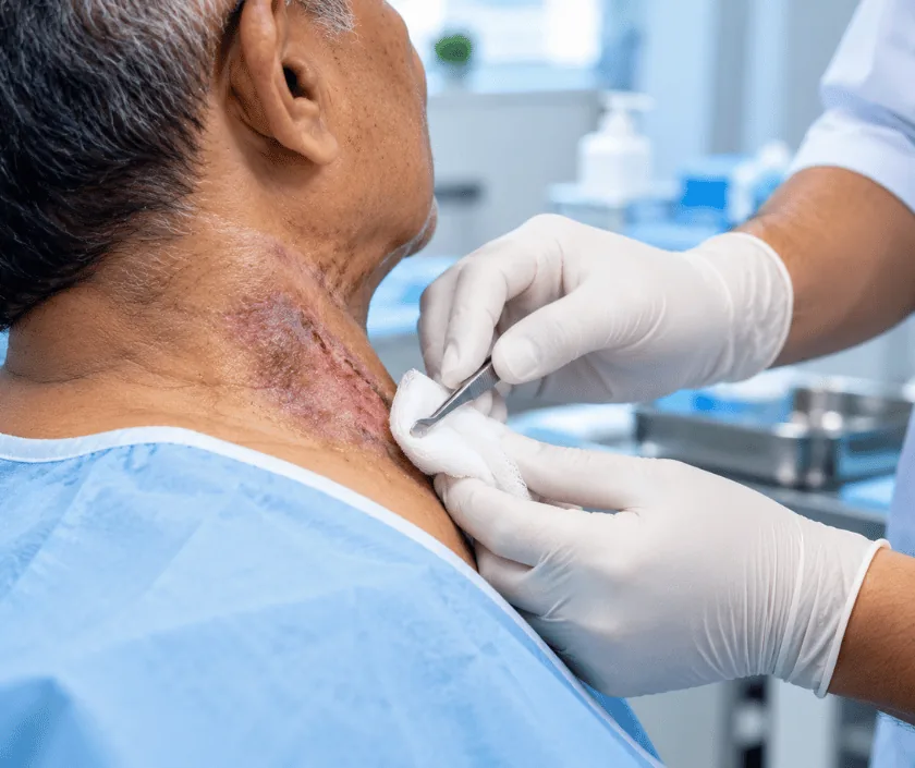
A radiation ulcer is a wound in skin that was treated with radiotherapy in the past. Radiation thickens and narrows the small blood vessels in that area, so the tissue never regains a normal blood supply. Wounds there heal poorly, and closure usually needs healthy tissue moved in from outside the treated area.
Radiotherapy works by damaging cells that divide quickly. Tumour cells suffer most, which is the point of the treatment, and healthy tissue in the beam recovers to a degree. Small blood vessels in that area, though, are altered lastingly. Their walls thicken, their channels narrow, and the tissue they feed is left with less circulation than before.
Years may pass with no trouble at all. Then a minor knock, a biopsy or a small operation opens the skin, and the wound simply does not close. Edges look pale and stiff, surrounding skin feels leathery, and ordinary dressings make no difference because the problem lies in the blood supply rather than on the surface.
Two things follow. Firstly, any tissue used to close the wound has to come from outside the irradiated field, since tissue inside it cannot support healing. Secondly, a biopsy is usually taken, because a wound that has stayed open in an irradiated area for a long time can occasionally contain a new cancer. Both points are explained before surgery is planned.
Surgery is offered when healthy tissue is available to bring in and when the original illness is under control. Both conditions matter.
The wound is examined and photographed, the old treatment field is mapped and a biopsy is usually taken. Scans may be arranged to show how deep the damage extends.
Because irradiated tissue cannot be relied on, the surgeon chooses a donor area well outside the treated field. Vessels are checked before any operation is booked.
In theatre the ulcer and the surrounding damaged tissue are removed generously, since leaving irradiated edges behind is a common reason for later breakdown.
A flap is moved into the defect, often as a free flap joined to vessels under a microscope. The donor area is closed directly or grafted.
The flap is watched closely at first, and review continues afterwards for both the wound and the condition originally treated.
Rest, careful positioning and regular flap checks. Pain relief is given and the wound and drains are monitored. Movement of the area is limited at this stage.
Swelling settles slowly. Healing in irradiated skin takes longer than usual, so stitches often stay in place longer and dressings continue for a while.
The flap has usually settled and daily activity increases. Any breakdown at the edges is treated early, because irradiated skin remains fragile.
The flap softens and colour differences fade a little. Regular review continues, both for the reconstruction and for the condition originally treated.
Healing in irradiated areas is slower and less predictable than elsewhere, so patience is part of the plan. The flap usually differs in colour and texture from nearby skin, and edges sometimes need a small further procedure. What most patients notice first is the end of daily dressings and a reduction in pain. Outcomes can vary with the dose given years earlier and with general health.
Operating in previously irradiated tissue carries a higher chance of wound problems, and that is stated plainly before consent.
Irradiated skin stays fragile for a long time, so home care focuses on protecting the area and reporting problems early.
Skin may look settled, yet the small vessels stay narrowed for years, which is why wounds there still heal poorly.
Grafts need a well supplied bed. Irradiated tissue rarely provides one, so a flap from outside the treated area is used.
Not usually, though a biopsy is taken because an old ulcer in a treated field can occasionally contain a new tumour.
Reconstruction is often possible many years later, provided healthy donor tissue is available and the original illness is controlled.
Elegance Clinic in Surat plans these operations with the full treatment history to hand, so the old radiotherapy field, the biopsy result and the reconstruction are considered together rather than separately.

Cost reflects the size of the area removed, the flap chosen and the longer healing these wounds usually need. Free tissue transfer requires microsurgery and closer monitoring, so it is estimated differently from a local flap. A written estimate is prepared after assessment and includes surgery, anaesthesia, stay, biopsy and follow up dressings. Where treatment relates to earlier cancer care, insurance cover is checked against the policy in advance.
These are the questions that come up most often in consultation. If yours is not here, send it on WhatsApp and the team will reply, usually the same day.
Ask your question →Cost depends on how much tissue must be removed, which flap is used and how long healing takes. A written estimate is prepared after assessment and includes surgery, the stay, biopsy and the dressings these wounds usually need afterwards.
It is done regularly, though wound problems are more common there than elsewhere. That is why healthy tissue is brought in from outside the treated field and why damaged edges are removed generously at surgery.
Longer than for an ordinary wound. Stitches often stay in for extra time and dressings continue for several weeks. Recovery can vary with the dose given years earlier, the site involved and your general health.
Most reconstructions do stay closed, although small breakdowns at the edges happen because surrounding skin remains fragile. These are usually treated with minor procedures rather than by repeating the whole operation.
It is not usually, but a wound open for a long time in a treated area can occasionally contain a new tumour. A biopsy is therefore taken during assessment, and removed tissue is examined as well.
There is no fixed pattern. Some ulcers appear within months, others many years afterwards, sometimes triggered by a small injury or a minor operation in the treated area. Any wound there that stays open deserves review.
Bring your radiotherapy records, the treatment site details, any oncology letters, previous biopsy reports and your medicines. The wound is examined and photographed, and the plan and estimate are explained before anything is booked.
Each technique below has its own page explaining how it works and when it is chosen.
Seeing patients from across the city and beyond: read about the practice on the plastic surgeon in Surat page, or check what a written estimate covers on the costs and insurance page.
