An arteriovenous malformation, shortened to AVM, is a direct connection between arteries and veins that forms before birth. This page explains why it is the most demanding of the vascular malformations, how it is treated and what to expect.
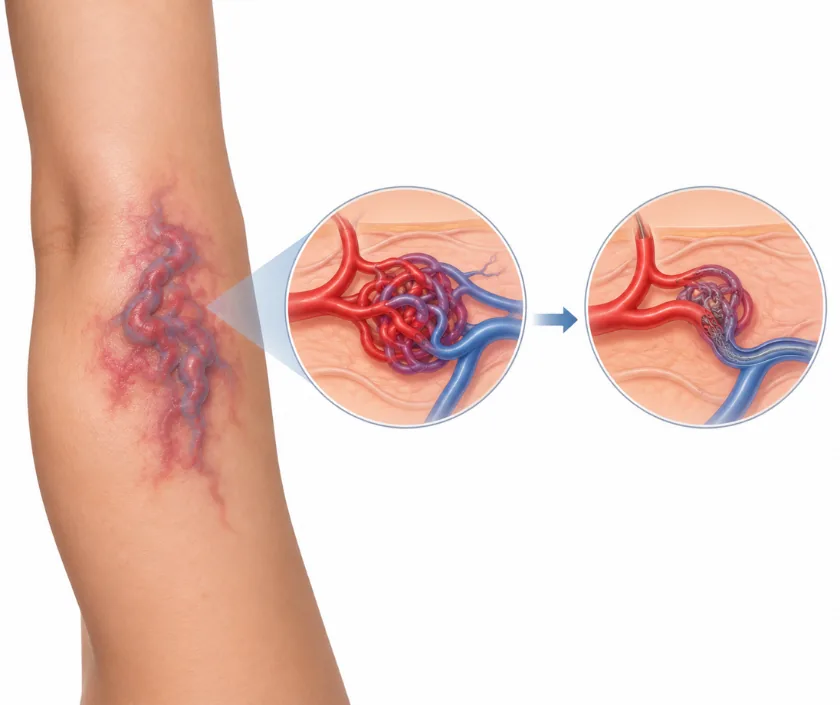
An arteriovenous malformation is an abnormal direct link between arteries and veins, without the tiny vessels that normally sit between them. It is present from birth, does not fade, and can enlarge at puberty, in pregnancy, or after injury or incomplete treatment. Care usually combines blocking the abnormal vessels with surgery, and long term review matters because the lesion can return.
Blood normally passes from arteries into tiny capillaries before it reaches the veins. In an arteriovenous malformation those small vessels are missing, so blood rushes straight through. The area feels warm, may pulse under the fingers, and can look like a pink patch long before anything else is noticed.
This is a malformation, so it does not fade the way an infantile hemangioma does. It grows with the child and can enlarge sharply at puberty, during pregnancy, after an injury, or after treatment that removed only part of it. Over years it may cause pain, bleeding, skin breakdown or strain on the heart when it is large.
Assessment uses ultrasound with Doppler, MRI and often an angiogram, a scan of the vessels showing exactly where blood enters and leaves. Treatment usually combines embolisation, where the feeding vessels are blocked from the inside, with surgical removal soon afterwards. Planning is done by a team, and honest discussion about what can be achieved comes first.
Scans are done first to map where the feeding vessels come from and how far the tangle spreads. Treatment is offered when the malformation is causing symptoms or is clearly growing.
Ultrasound with Doppler shows fast flow, and MRI maps the extent. An angiogram outlines the feeding arteries and the draining veins, which is essential before any treatment can be planned properly.
A surgeon and an interventional radiologist decide together what is realistic. For some lesions the aim is complete removal, while for others it is control of symptoms, and that is said plainly.
Through a fine tube placed in an artery, material is delivered to block the abnormal connections. This reduces blood flow and bleeding, and is usually timed shortly before the operation rather than used alone.
The lesion and the abnormal tissue around it are removed, and the defect is closed with local tissue, a flap or a graft. Blood may be needed, and larger removals are sometimes staged.
Because these lesions can return, reviews continue for years with scans. Regrowth found early is easier to treat than a lesion that has quietly enlarged again over a long period.
Pain, swelling and bruising are expected, and hospital care may last several days. Drains, dressings and careful watching of the wound and the blood supply are part of that time.
Wounds heal and dressings reduce. Activity builds slowly, and any flap or graft is checked at each visit. Sport and heavy work stay on hold.
A review with scans checks how much of the lesion has gone. Scars are still firm and red, and further stages are planned if the original plan was staged.
Regular review continues, because regrowth is possible even years later. Any new warmth, throbbing or swelling should bring the next appointment forward.
The honest picture is control rather than a one time fix. Blocking the feeding vessels and removing the tangle usually settles symptoms well, but the body can open new channels over months or years, so repeat treatment is common. Complete removal is possible for small, well mapped malformations. Where the tangle wraps around nerves or important structures, the aim shifts to keeping it quiet and protecting function. Regular scans and reviews continue for many years.
This is the most difficult of the vascular malformations to treat, and outcomes are less predictable than for the others. Risks and the chance of return are discussed frankly before any plan is agreed.
Watch the area, protect it from knocks, and know what a warning sign looks like.
Malformations do not fade or disappear. Infantile haemangiomas are the ones that usually shrink over years, and they are a different condition.
Partial treatment can make an arteriovenous malformation recruit new feeding vessels and become more active.
These need long review, because flow can return through new channels months or years later.
Laser works on some surface vessel conditions. A deep high flow tangle needs mapping, blocking of feeding vessels and usually surgery.
Families want someone who will say plainly what treatment can and cannot achieve with a condition that needs following for years.

Cost depends heavily on the size and site of the lesion, the imaging required, whether embolisation and surgery are both needed, the materials used to block the vessels, theatre time, blood products and the length of stay. Staged treatment multiplies these. Review scans continue for years and form part of the plan. A written estimate is prepared after assessment.
These are the questions that come up most often in consultation. If yours is not here, send it on WhatsApp and the team will reply, usually the same day.
Ask your question →No standard band exists, since treatment ranges from a single embolisation to staged surgery with reconstruction. Imaging, embolisation materials, theatre time, blood products and hospital stay all count. A written estimate is prepared after assessment, and insurance cover is usually discussed at the same visit.
Yes, and this needs saying clearly. These lesions can grow again months or years later, particularly when only part was treated or when embolisation was used on its own. That is why long term review with scans continues even when the result looks good.
Main concerns are bleeding during surgery, damage to nearby nerves and skin loss over the treated area. Blood may be needed. A team plans imaging and embolisation before the operation to reduce blood flow, which makes the surgery safer than it would otherwise be.
Expect several days in hospital after a significant removal, with swelling and bruising for two to three weeks. Wounds settle over about six weeks, and flaps or grafts are watched closely. Sport and heavy work wait longer, guided by the site treated.
Usually not on its own. Blocking the feeding vessels reduces flow, but the abnormal connections often recruit new supply and the lesion rebuilds. Embolisation is therefore most useful shortly before surgery, or to control bleeding and pain when removal is not safe.
Treatment is guided by symptoms and growth rather than by age alone. Bleeding, skin breakdown, pain or rapid enlargement move it forward. A quiet lesion may be watched, but it should not be ignored, since these lesions tend to progress over years.
The history and the pattern of growth are discussed, and the area is examined for warmth and pulsation. An ultrasound with Doppler is usually done, with MRI and angiography arranged. You will hear what is realistic, including whether control rather than removal is the aim.
Seeing patients from across the city and beyond: read about the practice on the plastic surgeon in Surat page, or check what a written estimate covers on the costs and insurance page.
