Tessier clefts are rare splits that run across the face in set lines, affecting the eyelid, cheek, nose or jaw. This page explains how they are numbered, how care is staged in Surat and what to expect at each step.
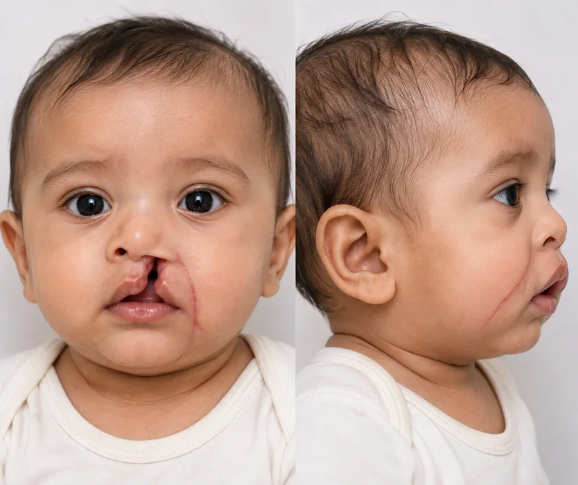
Tessier facial clefts are rare gaps in the soft tissue and bone of the face that follow set lines running out from the mouth, nose and eye. Surgeons describe them by number, so teams everywhere use the same language. Treatment is staged across childhood and matched to the structures each cleft involves.
Facial clefts form when parts of the developing face fail to fuse. Unlike a common cleft lip, these splits can run up through the eyelid and forehead or out towards the ear, and they involve bone as well as skin. Each child looks different, and two children with the same number can need quite different plans.
A numbering system is used to describe where a cleft runs. Lines are counted around the eye socket, outwards from the midline, with the number above the eye added to the number below to describe a full split. That shorthand tells any craniofacial team which structures are likely to be involved.
Care is arranged in stages over years. Protecting the eye and supporting feeding come first, then soft tissue closure, then bone work as the face grows. Ophthalmology, dentistry, speech therapy and paediatrics all sit alongside surgery, because these clefts rarely affect one structure alone.
Every child with a Tessier cleft needs assessment, but what is done first and when varies widely. The first plan is built around breathing, feeding and protecting the eye.
The face is examined and the cleft is described by number. Scans show which bones are missing and how the eye socket and jaw are affected, so nothing is discovered for the first time during surgery.
Eye protection, safe feeding and a clear airway come before appearance. Ointment, lid surgery or a feeding plan may all be arranged in the early months while the wider plan is drawn up.
Skin and muscle are brought together along the cleft line using local flaps. Careful planning keeps the scar in natural creases and restores the shape of the eyelid, nose or lip.
Missing bone in the socket, cheek or jaw is rebuilt with grafts or implants once growth allows. Planning software and models are used so the shape is worked out before the operating day.
The face keeps changing, so the plan is revisited at intervals. Further stages, dental treatment and refinements are added at points that suit school life as well as growth.
Recovery depends on the stage performed. Swelling is marked after facial surgery and is watched in hospital. Pain relief, careful wound care and gentle feeding are arranged before going home.
Swelling settles and stitches are removed or dissolve. School usually restarts once the wounds are sound. Contact sport and swimming wait longer, on the advice given for that stage.
Scars soften and the shape becomes clearer. Eye, speech and dental reviews continue. The next stage is planned only once healing from this one has fully settled.
Growth changes the face, so further operations are timed to it. Families are given a rough roadmap, which is updated at each review rather than fixed at the start.
These are among the more demanding differences in facial surgery, and the honest picture is one of steady improvement over years. Scars along the cleft lines remain visible. Bone grafts and soft tissue often need revision as the face grows, so most children have several operations before adulthood. Eye position, tear drainage and eyelid closure improve but may not become typical. Speech and hearing are followed alongside, since they are often affected too.
Rare facial clefts involve delicate structures such as the eye, the airway and growing bone. Risks vary with the stage planned, so each is discussed again before that operation.
Care at home changes with each stage. What stays constant is wound care, protecting the eye and keeping to the appointment schedule.
Tessier clefts follow different lines through soft tissue and bone, and often involve the eye socket, so the plan is quite different.
The face grows for many years. Rebuilding it works best in stages timed around that growth.
Scars are placed in natural lines wherever possible, but a visible mark along the cleft path remains.
Vision, tear drainage, hearing, speech and dental development are often involved, which is why a whole team is needed.
Rare facial clefts need a plan that spans childhood, so we set out the whole path with parents early rather than deciding one operation at a time.

No single band can be published for these clefts, because every child needs a different set of operations. Cost depends on which stage is planned, the operating time, implants or grafts used, hospital stay and the specialists involved. Scheme and insurance cover is assessed case by case, and staff help with the paperwork. A written estimate is given for each stage after assessment.
These are the questions that come up most often in consultation. If yours is not here, send it on WhatsApp and the team will reply, usually the same day.
Ask your question →No single band is published, because each child needs a different set of operations. Cost follows the stage planned, the operating time, any grafts or implants and the hospital stay. A written estimate is given for each stage once your child has been assessed.
Each stage is planned with a paediatric anaesthetic team and fitness is checked beforehand. Work near the eye and airway carries particular care, which is why scans and models are prepared in advance. Risks for that specific stage are explained before consent.
Recovery depends on the operation. Facial swelling is marked for the first week and settles over several weeks, and school usually restarts once wounds are sound. Each stage has its own timeline, which is given to you in writing.
Staged surgery can close the cleft, protect the eye, rebuild missing bone and give a much more balanced face. Some difference normally remains, since tissue is missing from birth. Aims are set stage by stage rather than promised in one go.
Function comes first, so eye protection, feeding and breathing are addressed in the early months. Soft tissue closure follows, and bone work is timed to growth. A rough roadmap is drawn at the first assessment and updated at every review.
Usually not, though difficulty breathing or an eye that cannot be protected does need same day attention. Poor feeding with weight loss also calls for prompt review. Seeing the cleft itself is frightening, yet the plan can be made calmly.
Your child is examined and the cleft is described by number. Scans and any earlier records are reviewed, photographs are taken and feeding, breathing and eye protection are checked. You will leave with a staged plan and a written estimate for the first step.
Seeing patients from across the city and beyond: read about the practice on the plastic surgeon in Surat page, or check what a written estimate covers on the costs and insurance page.
