When an ulcer reaches bone, the bone itself becomes infected, and antibiotics alone often cannot reach it. Surgery removes the infected part so the wound above can finally close and the rest of the foot is protected.
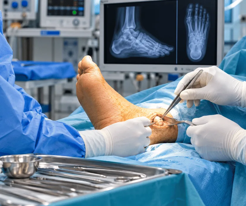
Foot osteomyelitis surgery removes bone that has become infected beneath a long standing ulcer. Antibiotics travel poorly into dead bone, so the affected portion is taken out, samples are sent for culture, and the soft tissue above is closed or covered. Clearing infected bone is often what finally lets the ulcer heal.
Foot bones sit very close to the skin. Under the ball of the foot, over a toe joint or at the heel, only a thin layer separates them from the outside world. When an ulcer deepens through that layer, bacteria reach the bone surface and then spread inside it. That is osteomyelitis, and it changes the whole treatment plan.
Diagnosis combines several findings. Probing gently to bone through a wound is a strong clue. Plain images can look normal early on, so scans are added when the picture is unclear. Blood tests help too, yet the most reliable answer comes from a bone sample taken during surgery and sent for culture, since surface swabs often grow bacteria that are simply passing through.
Treatment aims to remove what cannot recover. Infected and dead bone is taken out until healthy bleeding bone remains, and antibiotics then work on what is left behind. How much is removed depends on which bone is involved, and the surgeon balances clearing infection against keeping a foot that can still take weight.
Surgery suits a foot where infected bone can be removed while leaving a useful, weight bearing structure behind. Circulation and general fitness are assessed before any plan is made.
Probing, imaging and blood tests are combined to judge whether bone is involved. Narrowing of the arteries is looked for at the same time, since healing depends heavily on it.
During surgery a clean sample of bone is taken before antibiotics are given, so the laboratory can identify the organism accurately and a drug that actually works can be chosen.
Infected and dead bone is cut back until healthy bleeding bone is reached. Unhealthy soft tissue nearby is removed as well, and the wound is washed thoroughly.
Depending on the gap left behind, the wound is closed directly, kept open for later closure, or covered with a graft or flap once infection has clearly settled.
A tailored antibiotic course follows, guided by the culture. Weight is kept off the operated area, because pressure on a healing bone edge reopens the problem quickly.
Pain relief, sugar control and wound review begin at once. The foot stays elevated, antibiotics are adjusted when culture results arrive, and walking aids are arranged.
Dressings continue and stitches are checked. Antibiotics often carry on through this period, while the operated part of the foot stays free of weight.
Most wounds have healed and the antibiotic course is usually complete. Loading resumes gradually in a protective shoe, with the shape of the foot reviewed as you walk.
Attention shifts to preventing the next ulcer. Footwear, insoles and regular checks matter now, since removing a bone changes how pressure spreads across the sole.
Most wounds heal once the infected bone is gone and pressure is kept away, though the timeline can vary a great deal. The foot will be slightly different in shape, and that change alters how load travels through the sole. Because of this, a new pressure point can appear elsewhere and needs watching. Infection may also return if a small amount of affected bone was left behind.
These operations happen in a foot that already heals slowly, so the possible complications should be understood before consent is given.
Antibiotics and surgery only succeed when the foot is protected between visits.
Drugs reach dead bone poorly. Once infection is established inside bone, surgery is usually needed alongside them.
Surface swabs often grow bacteria that are only passing by. A bone sample taken in theatre is far more reliable.
Small, planned removals usually leave a working foot. Waiting instead often forces a much larger operation.
Nerve damage silences the warning, so bone infection can advance quietly under a wound that feels like nothing.
Elegance Clinic in Surat combines surgical clearance with careful microbiology, so antibiotics are chosen for the organism actually present. Dr. Ashutosh Shah plans how much bone to remove with the shape of the walking foot in mind.

Cost reflects how much bone is involved, whether more than one visit to theatre is needed and how the wound is finally covered. Laboratory tests, imaging and the antibiotic course add to the total as well. Admissions of this kind usually qualify under mediclaim, and a written estimate with insurance guidance is given before a date is booked.
These are the questions that come up most often in consultation. If yours is not here, send it on WhatsApp and the team will reply, usually the same day.
Ask your question →The figure depends on how much bone is involved, how many theatre visits are needed and the length of the antibiotic course. A written estimate follows examination, imaging and blood tests. Admissions like this are usually claimable under mediclaim, and cover is confirmed beforehand.
Removing infected tissue is often the safest thing to do, since leaving it allows the infection to spread. Sugar levels, kidney function and circulation are assessed first, and the anaesthetic is chosen to suit your general health.
Wounds commonly settle over several weeks while antibiotics continue. Weight stays off the operated area during that time, then returns in stages with a protective shoe. Recovery can vary with circulation, sugar control and how much bone was removed.
Return is possible, particularly if a small amount of affected bone remained or pressure falls on the same spot again. Culture guided antibiotics and proper offloading reduce that chance considerably. Regular reviews pick up any early sign of trouble.
Some early cases settle with a long antibiotic course when the bone is only superficially involved. Where bone is dead or exposed, drugs rarely reach it adequately. The options and their honest chances are discussed before you choose.
Ask for review whenever an ulcer stops improving, smells, discharges or exposes bone. Spreading redness, fever or a sudden rise in sugar means the same day. Bone infection treated early usually needs a smaller operation.
The wound is examined and gently probed, pulses and sensation are tested, and imaging is arranged. Blood tests and swabs are taken as required. Findings, likely surgery and a written estimate are then explained to you and your family.
Each technique below has its own page explaining how it works and when it is chosen.
Seeing patients from across the city and beyond: read about the practice on the plastic surgeon in Surat page, or check what a written estimate covers on the costs and insurance page.
