Tendons move by gliding inside a smooth sheath. Once that sheath is lost and the tendon lies open, it dries and sticks down, so cover is needed to protect both the tendon and the movement it controls.
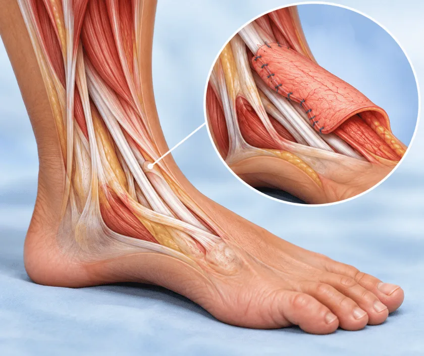
Exposed tendon coverage restores a soft, gliding layer over a tendon that has lost its sheath. Without cover the tendon dries, the surface dies and movement is lost as scar binds it down. Thin flaps of fascia, skin or muscle are used, chosen so the tendon can still slide beneath the new cover.
A tendon is a rope of collagen that transmits the pull of a muscle to a bone. It slides through a loose surrounding layer known as paratenon, which both feeds it and lets it move. When an injury removes skin and that layer together, the tendon lies bare. Air dries the surface within a day or two, and the outer fibres die.
Two problems follow. The tendon itself may fray or rupture, and scar tissue forms directly onto its surface, gluing it to whatever lies around it. Even if the wound eventually closes, the toe or ankle it controls may no longer move properly. That is why cover is planned promptly rather than watched for weeks.
The cover chosen has to be thin. A bulky muscle flap protects the tendon but can also limit gliding, so fascial flaps, thin skin flaps or a fascial free flap with a graft on top are often preferred. Where paratenon is still present, a skin graft may occasionally be enough, and the surgeon will look for that during assessment.
This surgery suits people with a clean wound over a tendon that is still intact enough to be worth protecting.
The wound is examined to see how much tendon is exposed, whether paratenon survives anywhere and whether the tendon still moves the joint it controls. Movement is tested where pain allows.
Dead skin, dried tendon surface and unhealthy tissue are removed under anaesthetic. The wound is washed and the true size of the defect becomes clear at this point.
A fascial flap, a thin local flap or a free fascial flap with a skin graft is selected. The aim is reliable cover that adds as little bulk as possible.
The flap is laid over the tendon and secured without tension, so nothing presses on the tendon or on the vessels feeding the flap.
A splint holds the ankle or foot in a safe position at first, and a therapy schedule is agreed for when gentle movement can begin.
The limb is elevated and the splint stays on. Flap colour is checked regularly. Pain relief keeps you comfortable enough to rest properly.
Dressings are reviewed and the graft, if one was used, is inspected. Gentle guided movement often begins under the therapist instructions.
Movement work increases and normal shoes may become possible. Swelling still varies with activity through the day.
Gliding continues to improve slowly. Scar massage and stretching remain useful, and stiffness that persists is reassessed at this stage.
Cover reliably protects the tendon, but movement depends on how much damage was already done and on the therapy that follows. Some stiffness is common, and full range is not always regained. The reconstructed area usually looks different in texture and colour from surrounding skin. Recovery can vary widely, so progress is judged over months rather than weeks.
Before agreeing to surgery it helps to know what can go wrong and how it would be handled.
Protecting the flap while gradually restoring movement is the balance to aim for in the first months.
Grafts need a living bed. Bare tendon without its surrounding layer will not support one, so a flap is generally required.
Total rest encourages scar to bind the tendon down. Guided movement at the right time is what preserves gliding.
Bulk can block the very movement being protected. Thin, pliable cover usually gives a better functional result.
Closure and movement are separate goals. Therapy after cover is what turns a healed wound into a working limb.
Tendon wounds are assessed with movement in mind, not just closure, so the plan is built around what the foot needs to do afterwards. Elegance Clinic explains that reasoning clearly at consultation.

The estimate reflects the type of cover chosen, the number of operations required, hospital stay and the therapy sessions that follow. A thin fascial free flap costs more than a small local flap, and the difference is explained at consultation.
Written estimates are prepared after assessment so the family knows what is included before admission. Where insurance applies, the office will guide you on the documents usually required.
These are the questions that come up most often in consultation. If yours is not here, send it on WhatsApp and the team will reply, usually the same day.
Ask your question →Fairly urgent. A tendon left open dries within days and the surface fibres die, which weakens it and encourages scar to bind it down. Prompt assessment lets the team decide whether cover can be done soon or whether the wound needs preparing first.
A written estimate is given after assessment. The figure depends on whether a local flap or a free flap is needed, how long the hospital stay is and how much therapy follows. Everything included is listed so the family can plan properly.
Movement usually improves but may not return completely. How much is regained depends on the original injury, how long the tendon was exposed and how consistently therapy is done. Honest expectations are discussed before surgery rather than afterwards.
Occasionally yes, when the loose layer around the tendon has survived and can nourish a graft. That is checked carefully at assessment. Where the layer is gone, a graft will not take and flap cover becomes the sensible option.
Many people manage ordinary footwear by around six weeks, though swelling often decides the timing. Soft, roomy shoes are usually easier at first. The team will advise based on how the flap and the wound edges are healing.
Discomfort is expected in the first days and is managed with regular pain relief. Most people find it settles quickly once swelling reduces. Elevation helps considerably, and the nursing team will adjust medication if pain is not controlled.
Bring any imaging, previous operation notes, discharge summaries and a list of current medicines. If dressings are in place, earlier photographs of the wound are helpful. Insurance details should also be carried so the estimate can be prepared accurately.
Each technique below has its own page explaining how it works and when it is chosen.
Seeing patients from across the city and beyond: read about the practice on the plastic surgeon in Surat page, or check what a written estimate covers on the costs and insurance page.
