The roof of the eye socket doubles as the floor of the front of the brain, so a fracture here concerns two teams at once. Assessment looks at the eye and at everything sitting above it.
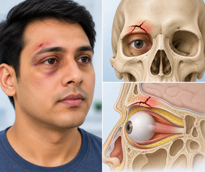
An orbital roof fracture is a break in the bone forming the top of the eye socket, which also separates the socket from the front of the brain. Many are managed without surgery once the eye is safe and no fluid is leaking. Operation is considered when fragments press on the eye, when the covering of the brain is torn or when the eye sits low.
Above each eye sits a thin curved plate of bone. On its underside it forms the roof of the socket, and on its upper surface it carries the front of the brain. Breaking it usually takes a heavy impact to the brow, a fall onto the forehead or a road crash, so this injury rarely arrives alone.
Signs tend to gather around the upper lid. Bruising is often dramatic, the lid may droop, and looking upwards can bring double vision if a fragment presses on the muscle above the eye. Should the eye be pushed downwards or forwards, that too is noticed within the first days. More concerning is any clear fluid from the nose, which suggests the covering of the brain has been torn where it lies against the bone.
Because of these overlaps, assessment runs on two tracks. An eye specialist checks vision, pupil reactions and movement, while the scan is reviewed with neurosurgical colleagues for any injury above the roof. Only then does a plan emerge, and for a good number of people that plan is careful observation rather than an operation.
Observation is the right answer for many roof fractures. Surgery is reserved for situations where the eye, the brain covering or the position of the eyeball will not settle on their own.
Vision, pupils, eye movement and lid position are recorded, and the CT scan is reviewed with neurosurgical colleagues. Whether the covering of the brain is intact shapes everything that follows.
Where the eye is safe and nothing is leaking, treatment is watchful. Repeat examination and imaging confirm that the fracture is stable and that the eye keeps its position.
An incision within the hairline allows the skin to be lifted forward, so the roof is reached from above without a scar on the face. Access is shared with neurosurgery when the brain is involved.
Fragments are lifted out of the socket and repositioned, and any tear in the covering of the brain is closed. Where bone is missing, the gap is bridged with a graft or a thin implant.
Vision is checked frequently through the first day and alertness is watched alongside it. Review then continues over the following months, and for longer in children whose skull is still growing.
Heavy bruising of the upper lid makes it hard to open the eye. Nursing staff check vision and alertness regularly, and the head is kept raised to help swelling drain.
Swelling falls enough for the lid to open properly and for eye movement to be tested. Quiet activity at home suits this stage, and reading may still be tiring.
Eye movement and lid position are formally reassessed. Return to work depends as much on the head injury as on the socket, so advice here is individual.
Any remaining double vision or difference in eye position is judged around now. Children stay under review for longer, since the fracture can change as the skull grows.
Vision is usually preserved, and most people regain comfortable single vision for everyday tasks. Double vision on looking far upwards can persist for a while and often improves slowly. Where the eye had been displaced, position generally improves although a small difference may remain. Because a head injury often accompanies this fracture, the pace of overall recovery is set by that as much as by the socket itself.
Any operation at the skull base carries particular risks, and these are set out plainly before a decision is made.
Care after this injury covers the eye and the head together, so instructions may come from more than one team.
Many roof fractures are stable and are watched rather than repaired. Surgery is chosen for pressure on the eye, a leak of fluid or a displaced eyeball.
Sight can be entirely normal while the covering of the brain is torn. Imaging and repeated examination are what settle the question, not how well you can read.
Young bone heals well, yet a fracture at the skull base can slowly enlarge as a child grows. That possibility is precisely why review continues for months.
Lid droop after this injury is usually caused by swelling and bruising around the muscle. It commonly improves as those settle, and it is measured at each review.
Injuries at the top of the eye socket are handled at Elegance Clinic in Surat with eye and neurosurgical colleagues involved from the first scan, so a decision to watch is made as carefully as a decision to operate.

What treatment costs depends on whether observation or surgery is chosen, whether a neurosurgical team shares the operation, the implants or grafts used and how long you remain in hospital. Since these follow from the scan and the eye assessment, an estimate is given in writing afterwards. Injury care of this kind is usually covered by health insurance and accident policies, and the team helps assemble the paperwork.
These are the questions that come up most often in consultation. If yours is not here, send it on WhatsApp and the team will reply, usually the same day.
Ask your question →The figure depends on whether surgery is needed, whether a neurosurgical team is involved, the grafts or implants used and the length of admission. A written estimate follows the scan and the eye assessment rather than preceding them.
Treatment after an injury is generally covered by health policies and accident cover, since it is not cosmetic. Emergency notes, imaging reports and photographs are usually needed for approval. Waiting periods and room rent limits in your policy still apply.
The operation is well described and is often shared with a neurosurgical colleague. Risks include fluid leak, infection, lid droop and double vision, and loss of vision is uncommon but possible. Each is explained before consent is taken.
Eyelid bruising settles across a fortnight, while overall recovery is usually set by the head injury alongside it. Many people return to light work in a few weeks. Recovery can vary considerably, so advice is given case by case.
Most people regain single vision for reading and for looking straight ahead. Some difficulty looking far upwards can remain and often improves slowly. Formal reassessment at six weeks and again at six months shows what is likely to last.
No. A stable fracture with normal vision and no fluid leak is watched with repeat examination and imaging. Surgery is advised for pressure on the eye, a torn covering of the brain or a displaced eyeball.
Vision, pupil reactions, eye movement and lid position are measured, and scans are repeated when needed. Children are followed for longer as the skull grows. Bring previous images and reports so changes can be compared properly.
Seeing patients from across the city and beyond: read about the practice on the plastic surgeon in Surat page, or check what a written estimate covers on the costs and insurance page.
