The eye socket is a cone of thin bone, and its shape decides where the eye sits. Reconstruction rebuilds the missing walls so the eye returns to its proper position and moves freely again.
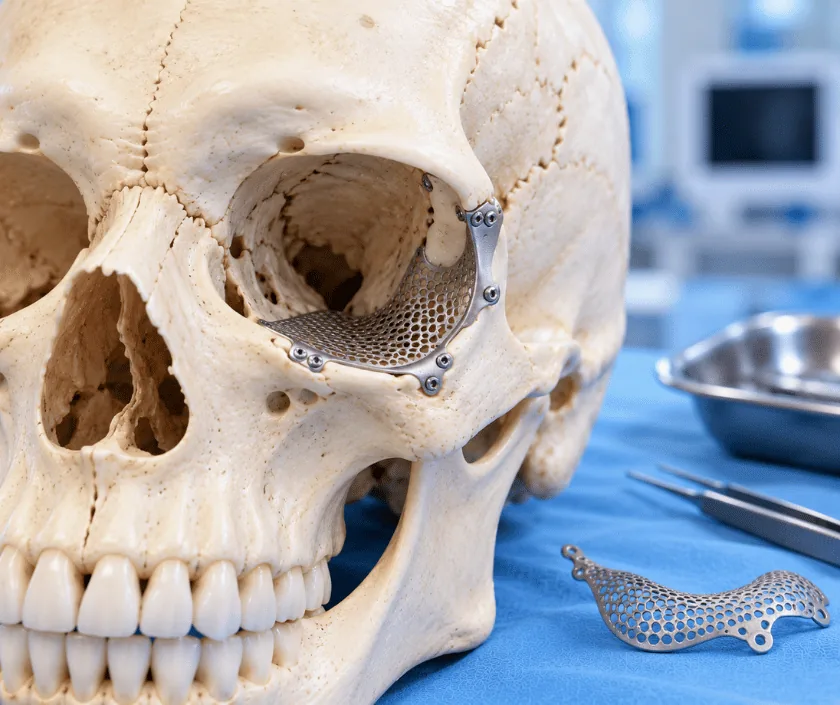
Orbital reconstruction rebuilds the walls of the eye socket when bone has been broken, removed or left to heal out of position. Thin implants, shaped from the scan, restore the volume of the socket so the eye can sit level with the other side. It is done at the time of an injury or later, when double vision or a sunken eye has not settled.
Think of the eye socket as a cone lined with bone and filled with the eyeball, its muscles, nerves and a cushion of fat. Everything in that space is held forward by the walls around it. Should one of those walls be broken outwards or lost altogether, the space grows larger, the contents settle backwards and the eye begins to look deeper set than its partner.
Reconstruction sets out to restore the container. A thin implant, most often a titanium mesh or a sheet of porous plastic, is shaped to follow the curve of the missing wall and placed on solid bone at the edges. Getting that curve right matters more than filling the hole, since the natural socket rises towards the back and a flat implant leaves the eye sitting too far in.
Planning comes from the scan. The uninjured side serves as a template, and the implant can be shaped in advance to match it. Where the socket has already healed in the wrong position, scarred tissue is released first, and that makes late surgery longer and less predictable than early repair.
Reconstruction suits people whose socket has lost its shape enough to alter eye position or eye movement. It is not helpful when the eye already sits correctly.
Vision, eye movement and the position of both eyes are measured, then the scan is studied. Comparing the injured socket with the normal side shows exactly how much volume has been lost.
A titanium mesh or porous sheet is contoured to match the missing wall, often before surgery begins. Following the natural rise towards the back of the socket is the part that decides the result.
Through a hidden incision in the eyelid or the eyebrow, the socket is opened and scar tissue is freed so displaced contents can return to their correct level.
The implant is laid on solid bone at the edges of the defect and fixed so it cannot move. Gentle movement of the eye by hand confirms nothing remains caught.
Eye position is compared with the other side on the table, wounds are closed and vision is tested as soon as you wake and repeatedly through the next day.
Swelling and bruising of the lids peak and vision is checked frequently. Sleeping propped up eases the swelling, and nose blowing is not allowed.
Bruising fades and the eye opens more easily. Double vision often changes during this stage as tissues settle, so it is measured rather than judged by feel.
Eye position and movement are formally reassessed. Most people are back at work, while swimming, dusty environments and contact sport still wait.
Position and any remaining double vision are considered settled around now. Further options, including prisms in glasses or muscle surgery, are only discussed at this point.
Eye position usually improves clearly, and many people find the difference between the two sides is no longer obvious to others. Exact matching is not the promise, particularly in late reconstruction where scarring limits how far tissue can be moved. Double vision often improves, though some people still need prisms in their glasses or a later muscle operation. Judging the outcome fairly takes about six months.
Rebuilding the socket is careful work in a confined space, and the risks below are set out before any decision is made.
The first weeks are about protecting the implant and keeping pressure away from the socket.
Nothing is done to the eyeball itself. The implant rebuilds the bony wall behind and beneath the eye, which is what supports it in the correct position.
Late reconstruction is possible and often worthwhile. It is more demanding than early repair because scar tissue has to be released, and the improvement may be less exact.
The amount of metal used is very small and sits inside the socket. It does not trigger security systems, and it is designed to stay in place without further attention.
Position improves a great deal, yet an exact match is not something any surgeon can offer. Honest measurement before and after surgery is what keeps expectations realistic.
Socket reconstruction at Elegance Clinic in Surat is planned by measuring the injured side against the normal one, and the aim of surgery is agreed with the patient before anything is booked.

The estimate reflects how many walls are rebuilt, the type of implant used, whether the socket has to be freed from scar tissue and how long you remain in hospital. Because these follow from measurement and the scan, the figure is given in writing after assessment. Reconstruction after injury or tumour surgery is usually covered by health insurance, and the team helps prepare the documents.
These are the questions that come up most often in consultation. If yours is not here, send it on WhatsApp and the team will reply, usually the same day.
Ask your question →It depends on how many walls need rebuilding, the implant selected, whether scar tissue has to be released and the length of admission. A written estimate is prepared once measurement and the scan are complete, so the figure matches your own situation.
Reconstruction after injury, infection or tumour surgery is medical treatment, so most health policies include it. Approval usually needs imaging, clinical notes and photographs. Waiting periods, room rent limits and exclusions written into your policy continue to apply.
It is a recognised procedure done under general anaesthesia, with vision checked repeatedly afterwards. Risks include infection around the implant, movement of the implant and lasting double vision. Loss of vision is rare, and frequent checking exists to catch problems early.
Swelling settles across a fortnight and many people return to light work within three to four weeks. Contact sport and swimming wait for clearance. Recovery can vary, especially when scar tissue from an older injury had to be released.
Position usually improves so much that the difference is no longer obvious to others. Exact matching is not something that can be assured, particularly in late reconstruction. Measurements before and after surgery keep the discussion honest.
Yes, and many people come years after the original injury. The socket is freed from scar tissue and the wall rebuilt, though the improvement is generally less exact than it would have been early on.
Vision, eye movement and the position of both eyes are measured, and the scan is reviewed with you. Bring old images, previous operation notes, insurance papers and a medicine list, plus photographs taken before the injury if you have them.
Seeing patients from across the city and beyond: read about the practice on the plastic surgeon in Surat page, or check what a written estimate covers on the costs and insurance page.
