Clearing a facial cancer safely often means removing more than the lump itself, which leaves a gap in skin, lining or bone. Post cancer facial reconstruction fills that gap with living tissue, so eating, speaking and appearance can be rebuilt.
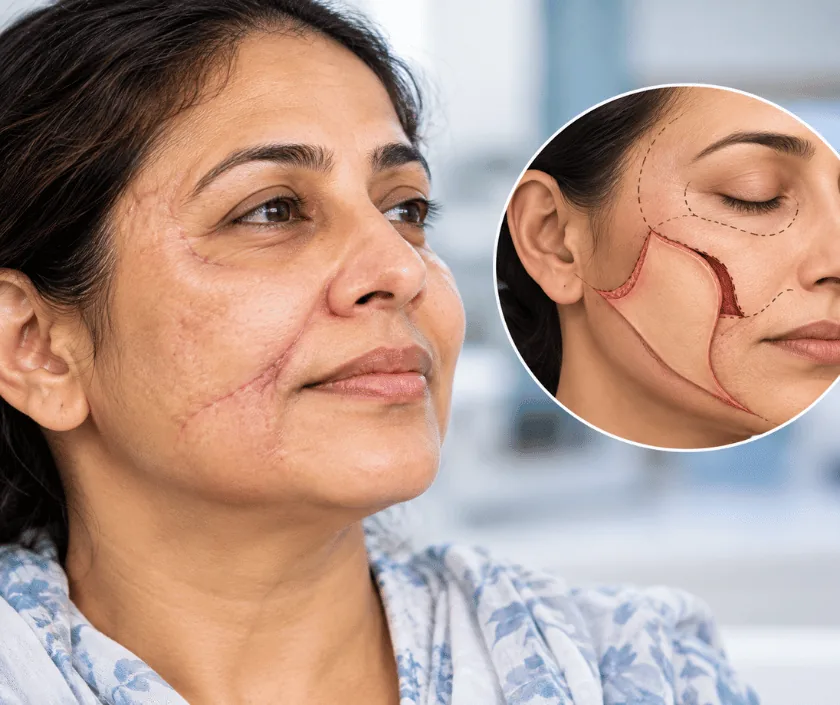
Post cancer facial reconstruction rebuilds the face after a tumour has been removed. The gap left behind may involve skin, muscle, lining of the mouth, bone or several of these together. Tissue is brought in from nearby or from another part of the body to restore shape and function. Reconstruction is usually done in the same sitting as the removal, so healing can begin at once.
Cancer surgery on the face has two jobs that pull in opposite directions. The tumour must be removed with a rim of healthy tissue around it, since anything left behind can grow again. That clearance, however, is what creates the defect, and the wider it needs to be, the more there is to rebuild. Planning therefore starts with the cancer surgeon and the reconstructive surgeon looking at the same scan together.
Once the extent of removal is known, the reconstruction is chosen to match. A small skin defect may close with a local flap of neighbouring skin. A hole through the cheek needs lining as well as an outer surface. When bone has gone, the replacement has to be bone, otherwise the face loses its support and the bite collapses. Tissue for this comes from the forearm, thigh, back or lower leg.
The face is also the part of us that other people read. Colour, texture and the position of the lip and eyelid all affect how a result feels to live with. Refinement of these details is normally left for a later sitting, once treatment is complete and tissues have settled.
The choice of reconstruction is made jointly with the cancer team, and it takes account of the defect, your general health and the treatment planned afterwards.
Cancer surgeon and reconstructive surgeon review the scans together and agree how much has to be removed. The reconstruction is designed around that plan, and donor sites are examined before the day of surgery.
The tumour is taken out with a margin of healthy tissue, and samples are checked during the operation where possible. Neck glands are removed at the same sitting when the disease requires it.
A second team lifts the chosen tissue with its blood vessels while removal is still under way. Working in parallel shortens the total operating time considerably for the patient.
Tissue is shaped into the defect and its vessels are joined to vessels in the neck under a microscope. Bone is contoured and plated so that the jaw line and bite are restored.
The flap is checked frequently in the first days, since early problems can often be rescued. Speech and swallowing therapy begins as soon as it is safe to start.
These days are spent under close observation, with the flap checked regularly. Feeding often goes through a fine tube at first, and swelling of the face and neck is expected.
Drains and tubes come out as things settle, and swallowing trials begin with the therapist. Many people move from hospital to home within this stretch.
Wounds have usually healed and diet has widened. If radiotherapy is planned, it commonly starts once healing allows, and the team explains what to expect from it.
Swelling continues to settle and the flap softens. Dental rehabilitation and any refinement of shape are usually considered from this point onwards.
Reconstruction restores shape and function, though it does not hide the fact that surgery took place. Flap skin usually differs in colour and texture from facial skin, scars are visible and some stiffness remains. Speech and swallowing improve with therapy, yet a few people continue to find certain foods difficult. Radiotherapy adds its own tightening. Progress can vary widely, so goals are reviewed honestly at each stage rather than promised in advance.
This is major surgery carried out alongside cancer treatment, and the risks deserve plain discussion before you consent.
Recovery continues well beyond discharge, and steady daily habits protect both the reconstruction and your general health.
Closing the defect with living tissue at the same sitting protects the area and allows radiotherapy to start on time. Delay usually makes the reconstruction harder, not easier.
Donor sites are chosen so that function is preserved. There is a scar and often some numbness or stiffness, but most people return to their usual activities.
Removing a tumour with a clear margin is the mainstay of treatment for many facial cancers. The evidence does not support the idea that operating causes it to spread.
Appearance is only part of it. Follow up visits, therapy, dental work and checks for recurrence all continue for years afterwards.
Reconstructive care at Elegance Clinic in Surat is planned alongside the cancer team, so removal, rebuilding and rehabilitation belong to one agreed sequence rather than three separate journeys.

Cost depends on the size of the defect, whether a free flap is needed, the length of the operation and the days spent in hospital afterwards. Implants, plates and dental rehabilitation are quoted separately when they are planned. Because cancer reconstruction is medically necessary, mediclaim policies and government schemes usually cover it, and a written estimate is prepared once the surgical plan is agreed with the oncology team.
These are the questions that come up most often in consultation. If yours is not here, send it on WhatsApp and the team will reply, usually the same day.
Ask your question →The total varies with the size of the defect, whether a free flap is used and how long the hospital stay runs. A written estimate is prepared once the surgical plan is agreed. Dental rehabilitation and later refinement are usually quoted as separate items.
Cancer reconstruction is medically necessary, so mediclaim policies and government schemes generally cover it. Approval needs the biopsy report, scan reports and a surgical plan. Sub limits on room rent and any waiting period in your policy will still apply, so check the details early.
Combining the two is standard practice for facial cancer, because a wound closed with living tissue heals better and allows further treatment to start on time. The operation is long, so fitness is assessed carefully with blood tests, scans and a physician review.
Most people spend several days in hospital with close flap monitoring, then continue recovering at home. Swelling settles over some weeks and therapy for speech and swallowing carries on longer. Radiotherapy, when planned, usually begins once the wounds have healed.
Shape and function are restored, but the result will not look untouched. Flap skin differs in colour and texture, scars remain visible and some stiffness is usual. Later refinement can improve contour once treatment is finished and tissues have settled.
Not always. A long operation demands reasonable heart and lung fitness, and healthy blood vessels at the donor and recipient sites. Where a flap is unwise, simpler options such as a local flap or a prosthesis are discussed instead, with their limits explained.
Bring the biopsy report, all scan films and reports, letters from the oncology team and a list of your medicines. If treatment has already started, bring the radiotherapy or chemotherapy schedule as well, since timing shapes the whole plan.
Each technique below has its own page explaining how it works and when it is chosen.
Seeing patients from across the city and beyond: read about the practice on the plastic surgeon in Surat page, or check what a written estimate covers on the costs and insurance page.
