Cheek bones, eye sockets and the upper jaw together form the midface, and losing part of it affects vision, breathing and the shape of the whole face. Midface reconstruction restores that support using the simplest method that will do the job.
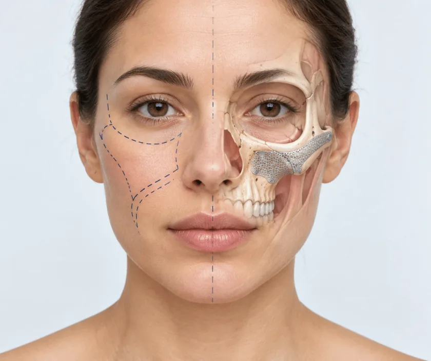
Midface reconstruction rebuilds the middle third of the face after injury, tumour surgery or a condition present from birth. Because this region supports the eye, separates the mouth from the sinus and carries the upper teeth, the goals are functional as well as cosmetic. Methods range from a simple prosthesis through bone grafts to free tissue transfer, chosen to match the size of the defect.
Think of the midface as a set of arches and buttresses that carry load from the teeth up to the skull. When part of that structure is lost, several things go wrong at once. The eye can drop or sink, the cheek flattens, the upper teeth lose their base and air passes between the mouth and the sinus, which affects speech and eating. Reconstruction has to answer all of those, not just the visible flattening.
Options are best thought of as a ladder. At the simplest level, a well made obturator, which is a removable plate that seals the defect, restores speech and swallowing without further surgery. Next come local flaps and non vascularised bone grafts, useful for small gaps with healthy tissue around them. Above that sit regional flaps that bring in muscle and skin from nearby, and at the top sits free tissue transfer, where bone and soft tissue are moved with their own blood supply and joined under a microscope.
Choosing between them depends on the size and site of the defect, whether radiotherapy is planned, the state of the remaining teeth and what the patient wants to manage day to day.
The right option depends on the defect and on the person. A frank discussion about daily management often decides the choice as much as the anatomy does.
A CT scan shows exactly which buttresses are missing and how the eye is supported. Dental models and photographs are added, and where useful a three dimensional model is printed for planning.
The team starts at the low end of the ladder and moves up only when needed. An obturator that works well may be preferable to surgery for a patient who is frail.
Where surgery is chosen, the buttresses are rebuilt with bone graft or with vascularised bone, held by plates. Orbital floor defects are supported with a plate or a thin implant.
Lining of the mouth and sinus is restored so that the two do not connect. Skin, muscle or fat is added to give the cheek back its fullness and contour.
Once healing is complete, an obturator, denture or implant supported teeth are planned with a dental colleague. This stage often decides how satisfied the patient finally feels.
Swelling of the cheek and around the eye is at its height. Feeding may be through a fine tube at first, and the reconstruction is checked frequently.
Swelling begins to fall and soft diet usually starts. Nasal and sinus care becomes part of the daily routine, and most people go home during this period.
Bone is settling and any temporary plate or obturator is reviewed. Eye position and vision are formally reassessed at around this point.
Contour becomes clear as swelling resolves. Dental rehabilitation and any refinement of the cheek shape are usually taken up from here.
Function usually improves a great deal, especially speech, swallowing and eye support. Appearance improves too, though the reconstructed cheek rarely matches the other side exactly and scars remain visible. Where radiotherapy has been given, tissues tighten and results are less predictable. Dental rehabilitation may take further months and is not always possible. Progress can vary widely, so each stage is reviewed on its own merits rather than promised at the outset.
Every rung of the reconstructive ladder carries its own risks, and these are explained for the option actually chosen.
Midface repairs sit next to the sinus and the mouth, so hygiene and gentle handling matter more here than almost anywhere else.
Bigger surgery is not automatically better. For many small defects a well made obturator restores speech and eating with far less risk, and it can be adjusted over time.
Restoring the orbital floor and the cheek support usually holds the eye in position. Where the eye has already dropped, correction is harder but often still worthwhile.
Sometimes they can, but often the bone must heal and settle first. Rushing this stage risks losing both the implants and the reconstruction underneath them.
Dental rehabilitation, contour refinement and long follow up are all part of midface care. Healing of the skin is an early milestone, not the finish line.
Midface work at Elegance Clinic in Surat is approached as a ladder of options, with the simplest workable choice discussed first and the reasons for moving up explained plainly.

Cost depends on which option is chosen. A prosthetic obturator sits at the lower end, bone grafting in the middle, and free tissue transfer with plates and implants at the upper end. Length of stay, intensive monitoring and any dental rehabilitation all add to the total. A written estimate is prepared after the scan and planning discussion, and reconstruction after injury or tumour is usually covered by insurance.
These are the questions that come up most often in consultation. If yours is not here, send it on WhatsApp and the team will reply, usually the same day.
Ask your question →The range is wide because the options are so different. An obturator costs far less than free tissue transfer with plates and implants. A written estimate is prepared once the scan is reviewed and the plan agreed, with dental work quoted separately.
Reconstruction after tumour surgery, injury or infection is medically necessary and usually covered by mediclaim policies and government schemes. Approval needs scan reports, the surgical plan and the underlying diagnosis. Room rent limits and policy waiting periods still apply.
These procedures are established and generally well tolerated, though the larger options are long and demand good heart and lung fitness. Risks include flap failure, infection and change in eye position. Fitness is assessed with blood tests, scans and a physician review beforehand.
Most people leave hospital within a few days to about two weeks depending on the option used. Swelling settles over some weeks, and the final contour is usually clear by around six months. Dental rehabilitation extends beyond that.
A proper seal between mouth and sinus usually restores clear speech and comfortable eating. Some people still find certain foods awkward, especially after radiotherapy. Speech therapy and a well fitted appliance make a considerable difference in practice.
Often yes, though it is a separate later stage. Bone must heal and settle before implants are considered, and radiotherapy can limit the options. A denture or obturator remains a reliable alternative when implants are not advisable.
Bring all scan films and reports, any biopsy result, previous operation notes and your current obturator or denture if you use one. A list of medicines helps, and so does writing down the problems that trouble you most.
Each technique below has its own page explaining how it works and when it is chosen.
Seeing patients from across the city and beyond: read about the practice on the plastic surgeon in Surat page, or check what a written estimate covers on the costs and insurance page.
