In a degloving injury the skin is peeled off the tissue beneath, often by a wheel or a roller, and it loses its blood supply while still looking attached. Treatment aims to keep what can survive and to replace what cannot.
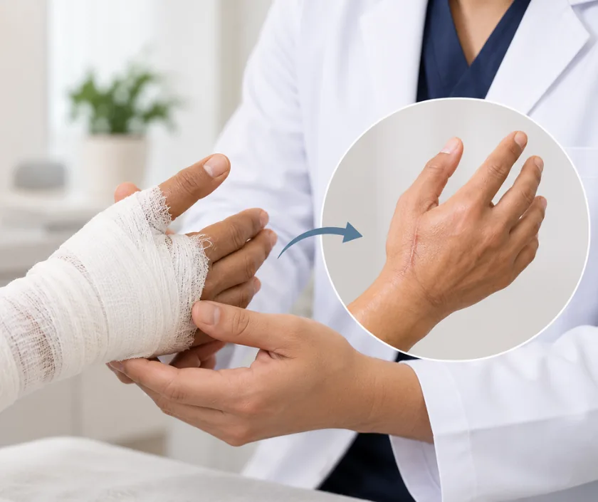
Degloving injury reconstruction treats skin that has been torn away from the tissue beneath it, usually by a rolling or crushing force. Even skin that looks intact may have lost its blood supply, so surgery removes what cannot survive, reuses what can, and covers the raw area with grafts or flaps in planned stages.
Picture a glove pulled off a hand. A degloving injury does something similar to skin, tearing it away from the layer that feeds it. It happens when a limb is caught between a wheel and the road, dragged along a surface, or pulled into a roller or a belt. Sometimes the skin comes right off. More often it stays attached along one edge, looking deceptively healthy while the vessels that supply it have already been sheared apart.
Judging what will survive is the central problem. Skin that seems pink on the day may turn dark within a few days, so wounds are inspected repeatedly and dead tissue is removed as it declares itself. Where the detached skin cannot survive as it is, it is often harvested, thinned and replaced as a graft on the same limb, which spares a separate donor site.
Once the area is clean, cover is restored with grafts, or with flaps that bring their own blood supply where bone, tendon or joint lies exposed. Movement and therapy run alongside, since a limb held still while it heals stiffens quickly.
Anyone with skin sheared off a limb, the scalp or the trunk needs this kind of staged surgery. What differs is the number of procedures and whether a graft or a flap gives the safer cover.
Circulation, bone injury and other wounds are checked first, because degloving rarely happens alone. Tetanus protection is reviewed and the limb is splinted before detailed planning begins.
Under anaesthesia the wound is washed out thoroughly and clearly dead tissue is removed. Doubtful areas are left for review, since the boundary between living and dead tissue often shifts over a few days.
Where it is suitable, the detached skin is thinned into a graft and laid back on the limb. This gives cover of matching colour and avoids taking skin from another part of the body.
The wound is inspected again after a short interval. Once the bed is clean, cover is completed with grafts, or with a flap where tendon, bone or a joint is exposed.
The limb is splinted in a position that protects the repair while limiting stiffness. Therapy begins as soon as the cover is secure, and it continues well after discharge.
The limb is elevated and dressings are left undisturbed unless there is a concern. Pain relief, fluids and antibiotics are given, and circulation is checked frequently.
Grafts are assessed as dressings come down, and further procedures are arranged if patches have not taken. Gentle supervised movement usually begins during this period.
Cover is generally stable though still fragile. Therapy becomes more active, and pressure garments or silicone may be introduced for the healing areas.
Scars soften slowly and swelling settles. Function keeps improving with therapy, and further surgery for contour or tightness is considered once the tissue has matured.
Most limbs recover useful function, though appearance changes for good. Grafted areas stay different in colour and texture, and the limb may look thinner where fat was lost. Recovery can vary a great deal, since two similar injuries behave differently depending on the blood supply left behind. Stiffness, sensitivity and swelling often continue for months, and further surgery is not unusual. Therapy shapes the final result at least as much as the surgery does.
These are severe injuries, and the risks reflect the injury itself as much as the operations used to treat it.
Recovery continues at home for months, and how the limb is used and protected during that time shapes the outcome.
Attachment is not the same as blood supply. Skin sheared off its base can look normal for days and then die, which is why wounds are reviewed repeatedly.
Dead tissue often declares itself gradually. A planned second look prevents infection taking hold in tissue that seemed alive at the first operation.
Joints stiffen within weeks. Guided movement starts as soon as the cover allows, because stiffness is harder to treat than the wound itself.
Degloving is treated in stages by design, with cleaning, then cover, then later refinement. Knowing this from the start makes the road ahead easier to plan.
Degloving injuries at Elegance Clinic in Surat are managed as a planned sequence, with the second look, the choice of cover and the therapy programme all discussed at the outset.

Treatment of a degloving injury is emergency and reconstructive care, so health insurance policies and government schemes usually consider it once the injury is documented. Cost depends on how many procedures are needed, the length of stay, whether grafts or flaps are used and the therapy that follows. Because treatment is staged, the estimate is revised as the plan develops, and a written estimate is given before each planned admission.
These are the questions that come up most often in consultation. If yours is not here, send it on WhatsApp and the team will reply, usually the same day.
Ask your question →Cost depends on how many operations are needed, the length of hospital stay, whether grafts or flaps are used and how much therapy follows. Staged treatment means the estimate is revised as the plan develops, and a written estimate is given before each planned admission.
Degloving is an accident injury, so mediclaim policies and government schemes generally consider treatment. Approval needs the accident record, photographs, operation notes and hospital paperwork. Employer or road accident cover may also apply, and reporting deadlines matter, so inform the insurer early.
Dead tissue often declares itself over several days rather than at once. A planned second look allows tissue that has since died to be removed before infection sets in, and it lets cover be placed on a bed that is genuinely clean.
Hospital stay usually runs to several days or longer, and dressings continue after discharge. Recovery can vary widely, since function returns over months with therapy. Return to work depends on the limb involved and on the physical demands of the job.
Useful function returns for most people, though appearance changes for good in the grafted areas. Colour and texture differ, and the limb may look thinner. Stiffness and swelling often improve slowly, and further surgery is sometimes needed for tight scars.
Anyone with active infection, uncertain tissue survival or an unstable general condition has cover delayed until things settle. Continued smoking works against grafts and flaps. Reconstruction is timed by how the wound looks rather than by a fixed schedule.
Circulation, bones and other injuries are checked before the skin wound itself. Tetanus protection is reviewed, photographs are taken and the limb is splinted. The staged plan, the likely number of procedures and the therapy programme are explained in writing.
Seeing patients from across the city and beyond: read about the practice on the plastic surgeon in Surat page, or check what a written estimate covers on the costs and insurance page.
