Removing a tumour from the chest wall can leave a gap in skin, muscle and sometimes ribs. Reconstruction restores a stable, protective wall, so that breathing stays efficient, the organs beneath stay covered and the area heals well.
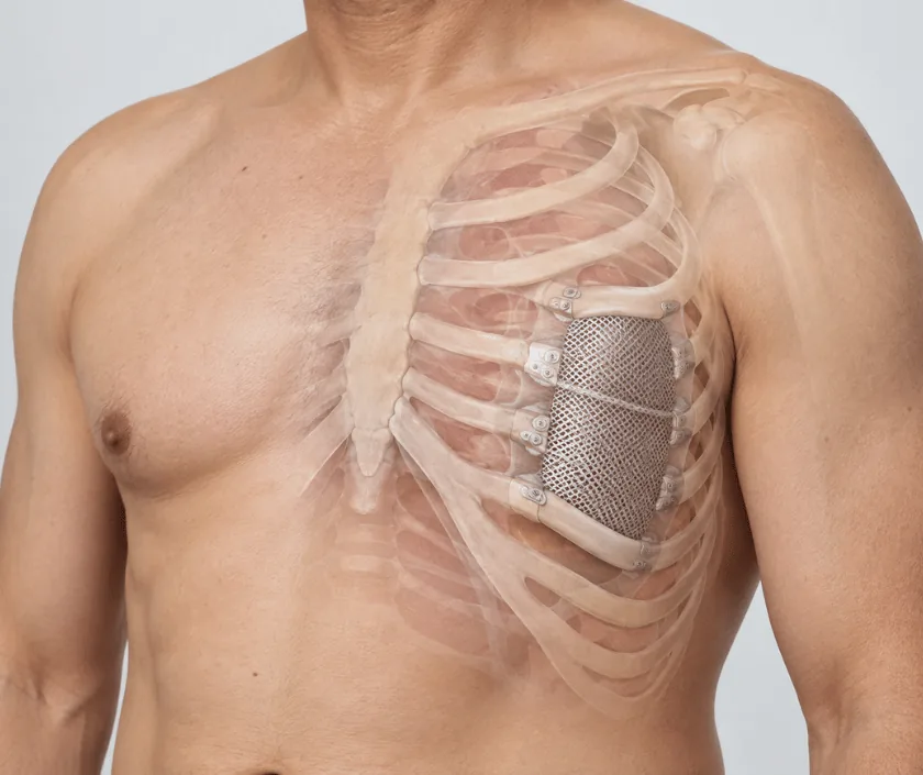
Chest wall tumour reconstruction rebuilds skin, muscle and bony support after a tumour is removed from the chest. When ribs are taken, a mesh or a rigid support holds the wall steady so that breathing is not affected. Muscle or a free flap then covers the repair with living, well supplied tissue.
Tumours of the chest wall arise from the ribs, from the soft tissue between them, or from skin and breast tissue that has grown inward. Some are sarcomas that start there, while others have spread from elsewhere or come back after earlier treatment. Removing them safely often means taking more than the lump itself, including a segment of rib and the lining beneath.
What is left behind decides the reconstruction. A small defect covered by nearby muscle needs nothing more. Once several ribs are gone, though, the wall becomes unstable, and a mesh or a rigid support is fixed to restore continuity. Without that support, the segment can sink inward on breathing in, which makes each breath less efficient.
Soft tissue cover comes next. Muscle from the back or the abdomen can be swung across on its own blood vessels, and a free flap is used when nearby options have been used up or damaged by radiation. Good cover matters here, because implanted mesh needs living tissue over it to stay free of infection.
Suitability turns on whether the tumour can be removed completely, and on whether the lungs and heart can handle a long operation.
Scans are reviewed with the oncologist and the thoracic surgeon, and the likely defect is mapped before the day of surgery. Donor options for muscle or a free flap are chosen at the same meeting.
The tumour comes out with a measured margin, which may include a segment of ribs and the lining beneath. Frozen section checks can be used during surgery to confirm the edges.
When several ribs are gone, a mesh or a rigid support is fixed to the cut rib ends. That layer holds the wall steady, so the segment does not move inward with each breath.
Muscle from the back or abdomen is swung across, or a free flap is brought in and joined to nearby vessels under a microscope. Living cover protects the mesh and helps the wound tolerate radiotherapy.
Drains remove air and fluid while the layers settle. Breathing is supported and watched closely, and physiotherapy starts early to keep the lungs clear.
Monitoring is close, with pain controlled well enough to allow deep breathing. Physiotherapy begins early, and drains stay in while fluid settles.
Drains come out as the output falls, and walking distance builds up gradually. Wound checks continue, especially over any implanted mesh.
Most daily activities return, though lifting and overhead work are still limited. Planned radiotherapy usually starts once the wound is sound.
Strength and stamina improve steadily. Scans and clinical checks continue with the oncology team, and shoulder movement is worked on when it remains stiff.
Most people end up with a chest wall that protects the organs beneath and moves reasonably with breathing. The area often feels firmer or numb, and a visible contour difference is common where ribs were removed. Recovery can vary, and stamina builds over months rather than weeks. Shoulder movement may need therapy when back muscle was used.
This is major surgery on a moving part of the body, so the risks deserve a frank discussion beforehand.
Breathing work sits at the centre of home care here, and everything else supports it.
Support with mesh or a rigid frame restores continuity, and living muscle cover holds it in place. Most people breathe normally once healing is complete.
Infection is a real risk, though covering the mesh with well supplied muscle reduces it greatly, which is why the flap forms part of the same operation.
Radiation makes tissue less forgiving. Bringing in a flap at the time of removal usually gives a wound that heals faster and tolerates treatment better.
Skin alone leaves no support and often breaks down over ribs or mesh. Layered repair with muscle or a free flap is what keeps the wall sound.
Chest wall work at Elegance Clinic in Surat is planned jointly with the oncology and thoracic teams, so removal, support and cover are decided as one operation.

Cost depends on the size of the defect, whether ribs and mesh are involved, the flap chosen and the intensive care needed afterwards. Implants and mesh are billed separately, and a longer stay adds to the total. Insurance often covers cancer surgery, and a written estimate is shared before admission.
These are the questions that come up most often in consultation. If yours is not here, send it on WhatsApp and the team will reply, usually the same day.
Ask your question →Free flap reconstruction usually falls between Rs 1.8L and Rs 4.5L, and the figure is case based. Mesh, rigid supports, theatre time and intensive care all shift it. A written estimate is prepared before admission and updated if the plan changes.
Breathing feels restricted for the first weeks while the wound settles. Once the wall is stable and the lungs are clear, most people breathe comfortably. Existing lung disease matters more here than the reconstruction itself.
Several days is typical, and longer when drains keep producing fluid or the flap needs close observation. Discharge depends on pain control, chest clearance and a settled wound rather than on a fixed number of days.
Mesh is widely used and generally well tolerated once healthy muscle covers it. Infection remains the main concern, which is why cover is planned in the same sitting and why fever is always taken seriously.
Usually yes. Living tissue cover is chosen partly for that reason, since it tolerates radiation better than a tight skin closure. Timing is agreed with the oncologist once the wound has healed sufficiently.
Lifting, pushing and overhead reaching are restricted for around six weeks, sometimes longer when back or abdominal muscle was moved. Physiotherapy guides the return, and heavy manual work needs a longer gap.
Bring all scans and reports, the biopsy result, records of earlier surgery or radiation and a list of medicines. Details of lung and heart conditions help, and a family member is useful during the discussion.
Each technique below has its own page explaining how it works and when it is chosen.
Seeing patients from across the city and beyond: read about the practice on the plastic surgeon in Surat page, or check what a written estimate covers on the costs and insurance page.
