The eyelid is thin, mobile and vital to the health of the eye. When a cancer is removed from it, reconstruction has to restore a smooth lid margin and a lid that still closes properly.
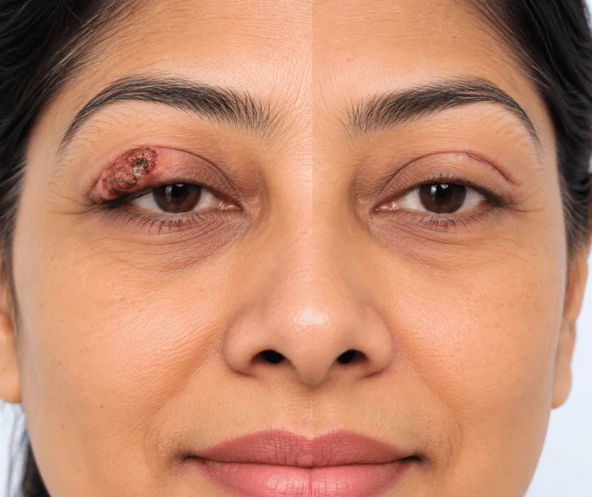
Eyelid cancer reconstruction is the repair done after a skin cancer has been taken off the eyelid. Because the lid protects and lubricates the eye, the surgeon rebuilds both the inner lining and the outer skin, and restores a smooth lid margin. Small defects close directly, while larger ones need tissue borrowed from the other lid or from nearby skin.
Skin cancer around the eye is common, and the lower lid and the inner corner are the usual sites. Removal has to take a margin of healthy tissue, so even a modest cancer can leave a defect that runs through the full thickness of the lid. That matters, because the lid is not simply skin. It carries a firm plate called the tarsus, a moist inner lining and a set of muscles that blink.
Reconstruction restores those layers in a way that keeps the eye protected. The surgeon checks how much of the lid width is missing, whether the margin itself is involved and whether the tear drainage passage at the inner corner has been affected. Each of those findings changes the plan.
Direct closure works well for narrow defects. Wider ones may take a graft of firm tissue for the inner layer with a skin flap over it, or tissue shared from the neighbouring lid. Where the tear duct is involved, a fine tube may be placed for a period so the passage stays open while healing.
Almost anyone who needs an eyelid cancer removed will also need some form of repair. What differs is the technique, and that follows the size and depth of the defect.
Vision, tear film and lid closure are checked along with the lesion itself. The surgeon measures how much lid width is involved and reviews any biopsy report before choosing a technique.
The cancer is taken with a rim of healthy tissue. The specimen is sent for examination, and reconstruction proceeds once the edges are reported as clear.
Firm tissue is used to replace the tarsal plate, so the lid keeps its shape and the inner surface stays smooth against the eye rather than rubbing it.
A local flap or a skin graft covers the outer layer. The lid margin is aligned carefully, because a step in that edge causes watering and irritation later.
Ointment, a light dressing and clear instructions follow. Reviews check lid closure, comfort and the position of the lash line as swelling settles.
Bruising and swelling around the eye are common and can look dramatic. Cool compresses, prescribed drops and rest with the head raised help most people through this stage.
Stitches are usually removed and vision feels clearer as swelling drops. Watering and mild grittiness often continue for a while and are expected.
The lid moves more freely and the scar is less firm. If a shared lid flap was used, the second stage to open the eye has usually happened.
The lid crease and lash line settle into their final position, redness fades and any small adjustment can be considered.
Most repaired lids look natural in everyday conversation, though the treated side may sit a little tighter or show a slightly different crease. Lashes do not grow back where the margin was removed. Mild watering can persist, especially after work near the inner corner. Swelling settles slowly, so judging the final appearance takes months rather than weeks.
Surgery this close to the eye is done carefully, but problems can still occur. Knowing them in advance makes the early recovery far less worrying.
Simple care protects both the repair and the eye itself. Written instructions and a contact number are given before you go home.
Many are, but a swelling that keeps returning at the same place, or that destroys lashes, needs a biopsy rather than repeated courses of ointment.
The operation works on the lid, not the eyeball. Vision is checked before and after, and protecting the surface of the eye guides the whole plan.
Skin replaces cover, not support. Without a firm inner layer the lid edge can turn or sag, so both layers are rebuilt when both are missing.
Lashes do not return where the margin was removed. Techniques aim to keep the lash line even, and many people find the difference hard to notice.
At Elegance Clinic in Surat, eyelid reconstruction is planned around the health of the eye first and appearance second, and Dr. Ashutosh Shah discusses each option and its trade offs before a date is fixed.

What an eyelid repair costs depends on how much of the lid is missing, whether the inner layer needs a graft and whether the tear drainage passage has to be addressed. Anaesthesia, theatre time, dressings and any tube placed at the inner corner all form part of the figure.
You receive a written estimate after the assessment, itemised so it can be checked against an insurance policy. Where the removal is part of cancer treatment, the team will explain which reports insurers usually ask to see.
These are the questions that come up most often in consultation. If yours is not here, send it on WhatsApp and the team will reply, usually the same day.
Ask your question →It varies with the size of the defect and the technique needed, since a narrow defect closes directly while a wider one needs grafts and flaps. A written, itemised estimate is prepared after the assessment so the family can plan and check insurance cover.
The work is on the lid rather than the eyeball, and vision is checked before and after. Risks such as watering, tightness or a lid that turns slightly are discussed openly, and most of these can be adjusted later if they persist.
Swelling and bruising around the eye peak in the first few days and can spread to the cheek. Most of it fades over the following couple of weeks. Mild puffiness in the morning may linger for longer and settles gradually.
The aim is a lid that works well and looks natural in conversation. Small differences in crease height, tightness or lash line are common. These usually become less noticeable as swelling settles over several months.
Often yes, though dryness is treated first and monitored closely, because a lid that does not close fully makes it worse. Your surgeon may choose a technique that favours lid closure over appearance in that situation.
When the margin is uncertain, waiting is safer than closing over it. A dressing is used for a short period and the repair follows once the edges are confirmed clear. This does not harm the final outcome.
Vision, lid closure and tear function are checked along with the lesion. Previous biopsy reports are reviewed, photographs are taken with consent, options are explained with their limits and a written estimate follows afterwards.
Each technique below has its own page explaining how it works and when it is chosen.
Seeing patients from across the city and beyond: read about the practice on the plastic surgeon in Surat page, or check what a written estimate covers on the costs and insurance page.
