The floor of the mouth is a small area that does a great deal of work, holding the tongue while letting it move freely. Reconstruction after cancer removal aims to keep that movement, so speech and swallowing are protected.
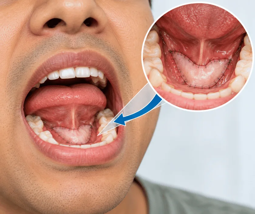
Floor of mouth reconstruction rebuilds the lining under the tongue after a cancer has been removed. Thin, pliable tissue is brought in as a flap so the tongue is not tethered to the gum. Skin grafts suit shallow defects, while deeper ones that involve the jaw or the tongue base usually need a free flap from the forearm or the thigh.
Under the tongue lies a narrow band of lining that separates the mouth from the muscles and glands of the neck. Cancer in this area is removed with a margin of healthy tissue, and because the space is tight, the resulting gap often reaches the gum on one side and the underside of the tongue on the other.
Closing that gap by stitching the tongue down to the gum is the problem to avoid. A tongue held in place cannot sweep food or shape sounds, so speech turns muffled and swallowing becomes slow and unsafe. Reconstruction instead lays in a fresh sheet of tissue that keeps a groove between tongue and gum. Shallow defects may take a skin graft. Deeper ones, or those that include part of the jaw, generally need a free flap with its own artery and vein joined to vessels in the neck.
Where the salivary ducts have been cut, they are usually redirected so saliva still drains into the mouth. If the jaw bone has been removed as well, bone reconstruction is planned in the same sitting.
This repair follows the cancer removal, so the decision is made jointly with the cancer surgeon. Your fitness for a long anaesthetic is assessed before anything is fixed.
Scans and biopsy reports are reviewed with the cancer surgeon. The likely gap is mapped, the donor site is picked, and dental review, nutrition support and chest checks are arranged before the date is fixed.
The tumour is removed with a margin of healthy tissue. Glands in the neck are usually cleared at the same time, which also brings the artery and vein for the flap into view.
A second team lifts a thin flap, often from the forearm, keeping its vessels attached. Thin tissue is chosen deliberately here, because bulk under the tongue restricts movement rather than helping it.
The flap is stitched in so a clear groove remains between tongue and gum. Vessels are joined under a microscope, salivary ducts are redirected, and the repair is checked for watertightness.
A temporary breathing tube and a feeding tube are commonly used at first, since swelling under the tongue is expected. Both come out once swelling eases and swallowing is tested safely.
Monitoring is close, with the flap checked regularly. The tongue is swollen and speech is limited, so a writing pad helps. Feeding is by tube and the mouth is rinsed gently as instructed.
Swelling settles and the breathing tube usually comes out. A swallow assessment guides the first sips and soft feeds. Discharge home often happens towards the end of this period.
Wounds are generally healed and diet has widened, though chewing may still be slow. Radiotherapy, if planned, often begins about now. Speech therapy continues for clarity of certain sounds.
The flap has softened and the groove under the tongue is established. Many people manage a fairly normal diet. Cancer surveillance visits continue on the schedule set by the oncology team.
Speech and swallowing tend to improve over several months, with the fastest gains in the first eight to twelve weeks. Taste and sensation under the tongue are often reduced and may stay that way. Some people manage a normal diet, while others keep certain foods off the menu for good. How far you get depends on how much tongue muscle was preserved, and radiotherapy can slow progress before it settles again.
The floor of the mouth sits close to the airway and the neck, so the specific risks here deserve plain description.
The routine at home is simple but needs consistency, particularly with mouth hygiene and swallowing practice.
Even a small removal here can tether the tongue if it is simply stitched. The size of the gap matters less than where it sits.
Under the tongue, bulk gets in the way. Thin, pliable tissue is chosen precisely so the tongue can still lift and move.
Early speech is affected by swelling and a temporary tube. Clarity usually improves over weeks and months, helped along by therapy.
A feeding tube is a bridge while swelling settles and swallowing is tested. Many patients return to oral feeding in stages.
The repair is planned with the oncology team so that removal and reconstruction happen in one properly organised sitting. Elegance Clinic in Surat explains the plan, the likely stay and the estimate before you are admitted.

The figure moves with the size of the removal, the type of flap, whether jaw bone is rebuilt, and how many days are needed in a monitored bed. Neck gland clearance in the same sitting is another factor.
An itemised written estimate is shared before admission. Many head and neck cancer operations attract insurance or government scheme cover, so bring your policy documents to the consultation.
These are the questions that come up most often in consultation. If yours is not here, send it on WhatsApp and the team will reply, usually the same day.
Ask your question →Free flap reconstruction generally falls in the band of Rs 1.8L to Rs 4.5L. The exact amount depends on the flap used, whether jaw bone is rebuilt and the length of hospital stay. A written estimate is provided before admission.
Not always, but it is common when swelling under the tongue is expected to be significant. It protects the airway during the first days and is usually removed within a week or two once the swelling has eased.
Feeding usually goes through a tube at first, then moves to sips and soft food once a swallow assessment shows it is safe. For many people this begins during the second week, although healing and radiotherapy plans can shift the timing.
Some sensation returns as nerves recover, but the reconstructed area is skin and rarely feels like the original lining. Taste often changes for a time, particularly during radiotherapy, and it may improve slowly over the months that follow.
Yes, though previously treated tissue heals more slowly and the risk of wound problems is higher. Free flaps are often preferred in that situation because they bring in tissue with a fresh blood supply from outside the treated field.
Surgery is usually arranged soon after the diagnosis and staging are complete, since delay allows the disease to progress. The date also depends on correcting nutrition, blood sugar and any chest problems first.
Bring biopsy reports, scans, a list of your medicines and any insurance papers. Bring a family member too, because the discussion covers the removal, the donor site, the likely stay and the recovery plan together.
Each technique below has its own page explaining how it works and when it is chosen.
Seeing patients from across the city and beyond: read about the practice on the plastic surgeon in Surat page, or check what a written estimate covers on the costs and insurance page.
