The upper jaw holds up the cheek, forms the floor of the eye socket and separates the mouth from the nose. Reconstruction after its removal restores that framework so the face keeps its shape and the mouth keeps working.
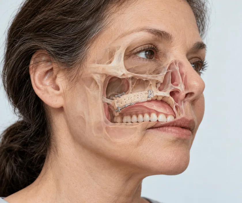
Maxillectomy reconstruction rebuilds the upper jaw after part or all of it has been removed for cancer. Depending on how much bone is taken, the surgeon may use a dental plate called an obturator, a soft tissue free flap, or a flap that carries bone. The aims are to seal the mouth from the nose, support the cheek and eye, and allow eating and speech.
The upper jaw is a hollow box of bone in the middle of the face. Its base is the roof of the mouth, its inner wall is the side of the nose, its top is the floor of the eye socket and its outer surface gives the cheek its projection. Removing it for cancer therefore affects eating, breathing, appearance and sometimes vision, all in one go.
Reconstruction is graded to the defect. When only the lower part is removed and enough teeth remain, an obturator plate may seal the opening well. When more of the box is taken, soft tissue is needed to fill the cavity and keep the mouth separate from the nose. Where the floor of the eye socket or the cheek support has gone, bone is usually required, and a flap carrying bone from the leg, hip or shoulder blade is used with its vessels joined to vessels in the neck.
Because appearance, vision and dental function all depend on this framework, the plan is made with the cancer surgeon, the eye surgeon where relevant, and the dental team before the day of operation.
The size of the removal decides how much reconstruction is required. Your fitness for a long operation is assessed alongside the cancer plan.
Scans are reviewed with the cancer surgeon and, where the eye is involved, the eye surgeon. The likely defect is classified, the donor bone is selected and dental needs are discussed before admission.
The tumour is taken out with a margin, through incisions placed to keep facial scars as discreet as possible. How much bone goes is confirmed during the operation itself.
A second team raises the chosen flap, taking bone from the leg, hip or shoulder blade when structural support is needed, or soft tissue alone when the cavity simply needs filling.
Bone is shaped and fixed with small plates and screws to restore cheek projection and eye socket support. Soft tissue lines the mouth surface and separates it from the nose.
The artery and vein of the flap are joined to neck vessels under a microscope. Blood flow is confirmed, drains are placed and the face is closed in layers.
You are watched in a monitored bed with regular flap checks. Facial swelling is marked and the eye on that side may be puffy. Feeding is by tube and the head is kept raised.
Swelling starts to fall and drains come out. Feeding by mouth begins in stages once the repair inside the mouth is judged secure. Most patients are discharged towards the end of this period.
Facial contour becomes clearer as swelling settles further. Radiotherapy, if advised, often starts around now. Walking is comfortable again where leg bone was used as the donor site.
Bone has united and the shape of the cheek has settled. Dental implants may be considered from this stage. Cancer follow up continues on the schedule set by the oncology team.
Facial shape is usually much closer to normal than it would be without reconstruction, though slight asymmetry is common and swelling takes months to settle fully. Where the eye socket floor was rebuilt, eye position often improves, yet mild double vision can persist. Chewing on the reconstructed side improves considerably once dental work is completed. Numbness over the cheek and upper lip is frequent and may not fully recover.
This is major surgery in the middle of the face, so the risks include those of the flap and those specific to the eye and the sinus.
Home care focuses on the mouth, the eye and the donor site, with clear instructions given for each.
The upper jaw can be rebuilt with bone and soft tissue, and appearance and function are usually far better than leaving the cavity open.
A plate suits smaller defects with enough teeth to grip. Large removals need tissue and often bone to support the cheek and the eye.
Where bone has been reconstructed, implants may be planned later. Radiotherapy affects the timing, so the dental team advises on this.
Swelling in the midface settles slowly and can take many months. Any sudden increase or new pain does need a review.
Elegance Clinic in Surat plans midface reconstruction with the cancer, eye and dental teams before the operation rather than after it. The family is told what the stay, the donor site and the estimate involve in advance.

Cost depends on whether bone is reconstructed, which donor site is used, how long the operation takes and how many days are spent in a monitored bed. Cases involving the eye socket usually sit at the higher end.
A written estimate is prepared once the joint plan is settled. Dental rehabilitation is quoted separately. Bring your insurance or scheme papers, as cancer surgery is often covered.
These are the questions that come up most often in consultation. If yours is not here, send it on WhatsApp and the team will reply, usually the same day.
Ask your question →Free flap reconstruction usually falls between Rs 1.8L and Rs 4.5L. Cases needing bone and eye socket support sit higher within that band. Operating time and hospital stay both affect the figure, and a written estimate is given before admission.
Some change is expected, since bone and soft tissue have been removed. Reconstruction restores much of the cheek projection and facial shape, though mild asymmetry is common. Swelling settles slowly, so the final appearance takes several months to emerge.
When the floor of the eye socket is removed, the eye needs support rebuilt beneath it. Double vision or a change in eye position can occur. An eye surgeon is involved in planning whenever the socket forms part of the removal.
Most stays run from about one to two weeks, depending on the flap used and how quickly swelling settles. Feeding by mouth usually restarts before discharge, and the family is taught mouth care and donor site care beforehand.
Where bone has been reconstructed, implants may become possible after healing is complete, often several months later. Radiotherapy influences the timing and the approach, so the dental team assesses this before any implant is planned.
Surgery is usually arranged once staging is complete and any diabetes, nutrition or chest problems have been addressed. Delay is avoided because the disease can advance, and radiotherapy afterwards has its own preferred timing.
Scans and biopsy reports are reviewed, the face and mouth are examined, and the likely defect is explained. Donor site options, the expected stay and later dental plans are discussed with the family, followed by a written estimate.
Each technique below has its own page explaining how it works and when it is chosen.
Seeing patients from across the city and beyond: read about the practice on the plastic surgeon in Surat page, or check what a written estimate covers on the costs and insurance page.
