Anotia means a baby is born with no outer ear on that side. This page explains how anotia differs from microtia, why hearing is checked early, how an ear can be rebuilt in stages and what shapes the cost.
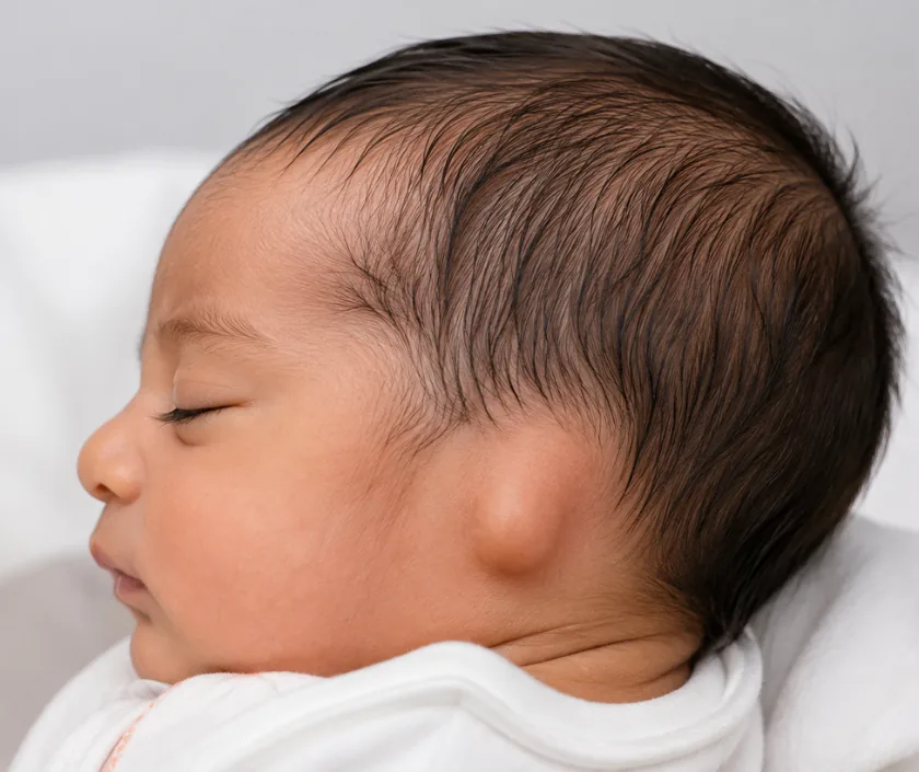
Anotia is the complete absence of the outer ear, usually with no ear canal on the same side. It differs from microtia, where a small folded remnant is still present. One side is affected far more often than both. Care starts with a hearing test, then the ear is rebuilt in stages using the child's own rib cartilage.
Anotia sits at the rarest end of a range of ear differences that a baby can be born with. In microtia the ear is small and incompletely formed, yet a fold of skin and cartilage is still there to work with. In anotia nothing of the outer ear has developed, so the side of the head is smooth, sometimes with a small skin tag where the ear would sit.
The outer ear and the ear canal form at the same time in early pregnancy. A child with anotia therefore usually has no canal on that side, and sound cannot travel the normal way. Hearing in the other ear is often normal, which is why speech usually develops on time. A formal hearing test is still arranged in the early months so that any support the child needs is in place well before school.
Rebuilding the ear follows the same path as microtia surgery. A framework is carved from the child's own rib cartilage, placed under a pocket of skin, then lifted away from the head at a later sitting. Our microtia reconstruction page sets out each stage in full.
The plan depends on the child age, the size of the chest, the quality of the skin over the area and what the family wants. Hearing is assessed first, whatever is decided about appearance.
A hearing test is arranged in the early months. When both ears are affected, a bone conduction hearing aid is fitted quickly so that sound reaches the brain while speech is developing.
A scan of the bone around the ear may be advised later in childhood to show whether a canal could safely be opened. The ear specialist and the plastic surgeon plan that question together.
Rib cartilage has to be large enough to carve a full sized framework. Chest size and general growth guide the date, so the first operation is usually planned in later childhood.
Cartilage is taken through a short cut low on the chest, carved into the shape of an ear and slipped into a pocket of skin. Gentle suction holds the skin against the fine detail.
At a later sitting the new ear is raised away from the head and a skin graft covers the back of it. Small touches may follow to balance shape, size and position.
Your child stays in hospital while pain from the chest and the ear is controlled. Drains and dressings are checked often, and deep breathing is encouraged so the chest stays clear.
Dressings are changed in clinic and the new ear is protected from knocks. Chest soreness settles steadily. Most children are back at school within two to three weeks.
The shape is reviewed and light activity returns. Contact sport, swimming and anything that presses on the ear wait until the surgeon is happy with healing.
Swelling settles and the outline becomes clearer. The next stage is booked once healing is stable, and further small adjustments may be discussed as your child grows.
A rebuilt ear looks like an ear and matches the other side closely from a normal talking distance, but the fine folds are softer than a natural ear and it stays slightly different on close inspection. It is built in stages, so the shape at the end of the first operation is not the finished result. The chest is sore for a couple of weeks. Building an ear does not by itself improve hearing.
Reconstruction for anotia is staged surgery on a growing child, so the plan is talked through in full before anyone commits. Knowing what can go wrong helps you raise a problem early.
Two areas need care at once, the new ear and the chest where the cartilage was taken.
Hearing depends on the canal and inner ear, not the outer ear shape. Many children hear well on the other side and are tested carefully to be sure.
Rib cartilage is usually not ready that early, and skin needs to be the right size. Waiting gives more material to build with.
It can match closely in size and position, but the fine detail stays a little softer than a natural ear.
These are separate problems with separate treatments, though they are often planned around each other.
Families value being given the honest choice between reconstruction and a custom made ear, rather than being pushed towards one of them.

Cost depends on how many stages are planned, the length of each operation, anaesthetic time, hospital nights and whether ear surgery for hearing is arranged alongside. Reconstruction for anotia follows the microtia pathway, so the published microtia band is the one that applies. A written estimate for each stage is given after your child is examined, and cover under a scheme or a mediclaim policy is checked before admission.
These are the questions that come up most often in consultation. If yours is not here, send it on WhatsApp and the team will reply, usually the same day.
Ask your question →The published band for microtia reconstruction runs from about Rs 1.4L to Rs 2.8L, and the same pathway is used for anotia. The final figure depends on the number of stages, theatre time and hospital nights. A written estimate is given for each stage before admission.
In microtia a small, folded ear is present but underdeveloped. In anotia no outer ear has formed at all, so the skin on that side of the head looks smooth. Surgery follows a similar route, though there is less of the original ear to build around.
Cartilage is taken through a short cut low on the chest, and the ribs carry on growing. Soreness there is expected for some weeks. Careful anaesthesia, good pain relief and breathing exercises form part of the plan, and children are watched closely on the ward.
Most children stay two or three nights, then rest at home. School often restarts within two to three weeks. Sport, swimming and anything that presses on the ear wait until the surgeon agrees, which is usually a couple of months.
The aim is an ear that matches in size and position and looks natural in photographs and under hair. A close match is realistic; an exact copy of the other side is not. Shape keeps improving over months as swelling settles.
Hearing on the affected side is usually reduced because the canal has not formed. When the other ear hears normally, speech generally develops on time. If both ears are involved, a bone conduction hearing aid is fitted early. An ear specialist guides this part.
The head and neck are examined, photographs and measurements are taken and any hearing reports are reviewed. You will hear how many stages are likely, roughly when they could begin and what each stage would cost, with the plan written down for you.
Each technique below has its own page explaining how it works and when it is chosen.
Seeing patients from across the city and beyond: read about the practice on the plastic surgeon in Surat page, or check what a written estimate covers on the costs and insurance page.
