When joints between the skull bones close too early, the head grows into an unusual shape. This page explains how craniosynostosis is assessed in Surat, when surgery is advised, what the operation involves and how children recover.
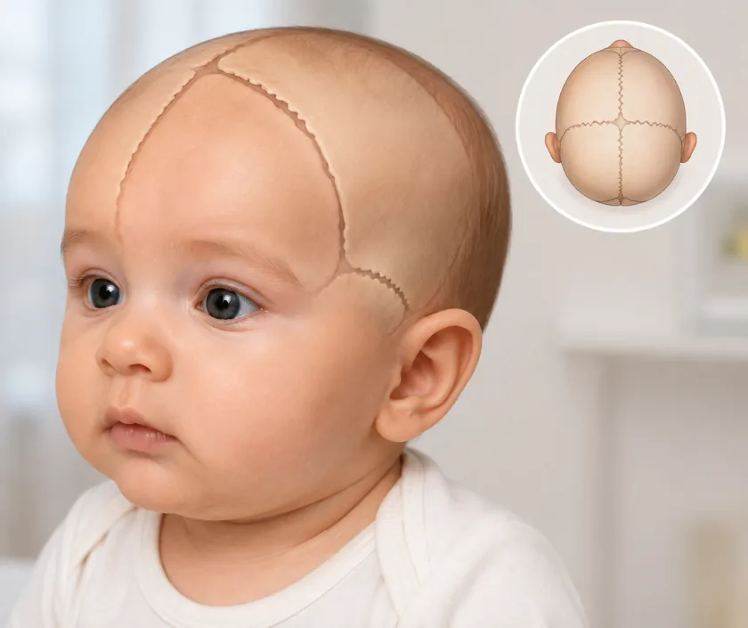
Craniosynostosis means one or more joints between the skull bones fuse too early, so the head grows in an unusual direction and pressure can build inside. Surgery opens the fused joint and reshapes the affected bones, allowing the brain room to grow. Assessment involves a paediatrician, scans and an eye check before any date is fixed.
A baby skull is made of separate plates joined by flexible seams. Those seams let the head grow quickly during the first years while the brain expands. If one seam fuses early, growth stops in that direction and continues elsewhere, which is why the head takes on a recognisable shape depending on which seam is involved.
Most babies with an odd head shape do not have this condition. Lying in one position flattens the back of the head in many healthy infants, and that improves with position changes and time. A true fusion is suspected from the shape, a ridge along the seam and the pattern of growth, then confirmed by examination and a scan.
Where a seam has truly fused, surgery aims to release it and reshape the skull so the brain has room. Eye checks and head measurements help judge whether pressure is rising. Operations are carried out in a hospital with paediatric intensive care, alongside a neurosurgical and anaesthetic team.
Surgery is planned by a craniofacial team after examination and imaging. The technique depends on which seam is involved and on how old the baby is.
Head shape and measurements are checked, the seams are felt and a paediatrician reviews development. An eye examination looks for signs of raised pressure inside the head.
A scan confirms which seams have fused and shows the shape of the skull in three dimensions. Models or planning software help work out the reshaping before the operating day.
Not every unusual head shape needs an operation. Surgery is advised when a seam is truly fused, when pressure may be rising or when the shape will not correct as the child grows.
Working with a neurosurgical team, the fused seam is opened and the affected bones are lifted, reshaped and fixed in a better position with fine plates or stitches that dissolve.
Children are watched in a high dependency or intensive care setting at first. Blood counts, fluid balance and swelling around the eyes are followed closely in the first days.
Your child is monitored closely and pain relief is given regularly. Swelling around the eyes is expected and often closes them for a time, which settles as the days pass.
Most children go home once feeding and comfort are steady. Swelling falls quickly in this period. Head wounds are kept clean and dry, and gentle activity is allowed.
The wound is reviewed and the head shape is measured again. Normal play is usually permitted. Advice is given about helmets, car seats and rough games.
Head growth, vision and development are followed for years. Small irregularities in the skull may show as hair grows, and further work is occasionally advised.
Head shape usually improves a great deal, and most children end up with a shape that draws no attention. Some unevenness generally remains and the two sides are rarely a match. The scar sits within the hair and becomes hard to see as hair grows over it. A small number of children need a further operation, particularly those with a syndrome affecting several seams. Reviews continue through childhood to check growth.
This is major surgery on a young child and is undertaken only where the assessment supports it. Being told the risks plainly helps parents weigh the decision properly.
Swelling of the face and eyelids in the first days is expected and can look alarming, yet it settles quickly.
A helmet guides growth but cannot open a seam that has fused. It is used after some operations, not instead of them.
Most unusual head shapes come from lying position and settle with repositioning. Examination tells the two apart.
It gives the growing brain room and relieves pressure. Appearance improves too, but it is not the only reason.
Skulls keep growing, so children stay under review for years to make sure growth continues as it should.
Families come to us for early assessment, a clear explanation of what the scan shows, and care planned with the craniofacial team around the baby.

No fixed band is published for skull surgery, because the operation, the hospital setting and the length of intensive care differ so much between children. Cost is shaped by operating time, the team involved, transfusion needs, plates or fixation used and the days spent in monitored care. Scheme and insurance cover is assessed individually. A written estimate is given after assessment.
These are the questions that come up most often in consultation. If yours is not here, send it on WhatsApp and the team will reply, usually the same day.
Ask your question →No fixed band is published, since the operation and the hospital setting vary widely. Operating time, the team involved, transfusion needs, fixation used and days in monitored care all shape the figure. A written estimate follows assessment, along with help on scheme cover.
It is major surgery and is only advised when the assessment supports it. Care is given in a hospital with paediatric intensive care and a neurosurgical team. Blood loss is the main concern, so transfusion is arranged in advance and monitoring stays close throughout.
Children usually spend several nights in hospital, some of it in monitored care. Swelling around the eyes peaks in the first days and settles within a fortnight. Most families are home inside two weeks, with a review at about six weeks.
Most children gain a much more even head shape and room for the brain to grow. Small irregularities can show as hair thins or partings change. Growth continues to alter the skull, so a further procedure is occasionally advised later.
Timing depends on which seam is fused, the shape and any sign of rising pressure. Many operations are planned in the first year, when bone remodels readily. A paediatric team gives an opinion before any date is chosen.
Flattening from lying in one position is common and usually improves with position changes and tummy time. True early fusion feels different, with a firm ridge and a shape that keeps worsening. A review settles the question rather than guesswork at home.
The head is measured and the seams are felt, development is reviewed and the eyes are examined. Scans may be arranged. You will hear whether the shape is positional or a true fusion, what surgery would involve and the written estimate.
Seeing patients from across the city and beyond: read about the practice on the plastic surgeon in Surat page, or check what a written estimate covers on the costs and insurance page.
