Bone that lies open to the air dries, dies at the surface and invites infection. Placing living tissue over it restores a protective layer and gives the bone underneath a chance to recover.
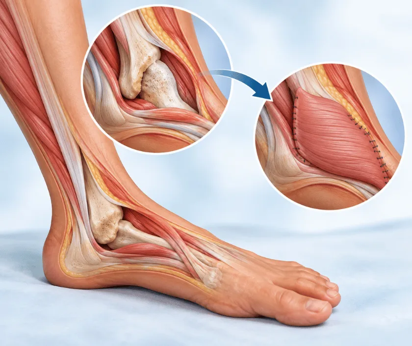
Exposed bone coverage is surgery that brings healthy tissue with its own blood supply over a patch of bare bone. Skin grafts rarely take on bone, so a flap is usually needed. The dead surface layer of bone is removed first, then muscle, fascia or skin is moved across and stitched into place.
Bone is living tissue, but its outer surface depends on a thin membrane called periosteum for blood supply. When an injury, a burn, pressure or an infection strips that membrane away, the surface of the bone is left without circulation. Within days it dries and the outer layer dies. Dressings can keep such a wound tidy for a while, yet they cannot make it heal.
Coverage surgery addresses both parts of the problem. First the dead bone surface is trimmed until healthy bleeding bone appears, and any infected tissue around it is removed. Then living tissue is brought across. Muscle flaps are often chosen when infection has been part of the picture, because muscle carries a generous blood supply. Fascia or skin flaps may suit shallower defects where bulk is not wanted.
The site decides much of the plan. Around the knee and upper shin, nearby muscle can often be rotated in. Lower down the leg the local options run out, so tissue is more often transferred from another part of the body and its vessels joined under a microscope.
Coverage works best when the underlying problem has been dealt with and the wound can be brought to a clean, stable state.
The wound is examined, the depth of bone involvement is judged and imaging is reviewed. Blood tests and sometimes a swab or bone sample help decide whether infection is still active.
Under anaesthetic the dead bone surface is trimmed until healthy bone bleeds, and unhealthy soft tissue around the defect is removed so the wound bed is clean.
The size of the defect and its position decide whether a local flap, a rotated muscle flap or a free tissue transfer will give reliable cover with acceptable donor site cost.
The flap is raised on its blood supply, moved into the defect and stitched in without tension. Free flaps need the artery and vein joined under magnification.
The area the tissue came from is closed directly where possible, or resurfaced with a skin graft, and is dressed and supported.
Rest and elevation are the priority. The flap is watched closely for colour and warmth, and pain relief is given regularly so you can stay comfortable and still.
Dressings are changed and stitches reviewed. Sitting out of bed and gentle movement usually begin. Most patients go home during this period with clear instructions.
The flap has usually settled and swelling is easing. Physiotherapy steps up and normal footwear becomes possible again for many people.
Bulk reduces slowly and the scar fades. Any refining procedure, if one is wanted, is generally discussed only after this point.
The realistic goal is durable, healed cover rather than an invisible repair. Flap tissue differs in colour and thickness from the skin around it, and it often looks raised at first. Softening happens over months. Sensation in the flap is usually reduced and may not return fully, so care around heat and pressure stays important in the long run.
Every method of covering bone carries risk, and knowing these in advance helps you judge whether to proceed.
The flap is fragile in the early weeks, so a few simple habits protect the work that has been done.
Bare bone without its membrane cannot do this. The wound may shrink at the edges but the centre stays open.
Medicines cannot reach dead bone. Surgical clearance and living cover are what change the situation.
A tidy dressing can sit over a wound that is going nowhere. Progress is judged by the wound bed, not the dressing.
The choice of flap matters. Tissue must reach without tension and carry a reliable blood supply to the exact spot.
Wounds with exposed bone are assessed carefully before anything is promised, and the plan is explained stage by stage. Elegance Clinic keeps the family informed about what each step is meant to achieve.

The final figure depends on the size of the defect, whether a local flap or a free flap is used, how many theatre visits are needed and how long you stay in hospital. Investigations and physiotherapy also form part of the total.
A written estimate is prepared after the wound has been assessed and any imaging reviewed. If insurance is involved, the team will explain which papers the insurer typically asks for and how approval usually proceeds.
These are the questions that come up most often in consultation. If yours is not here, send it on WhatsApp and the team will reply, usually the same day.
Ask your question →A graft survives by absorbing nourishment from the surface it is laid on. Bare bone that has lost its living membrane cannot provide that, so the graft simply dies. Tissue with its own blood supply is needed instead, which is what a flap provides.
A written estimate is given after assessment, because the figure changes with the type of flap chosen, the number of operations and the length of stay. Once the wound has been examined and imaging reviewed, the office prepares the estimate for you in writing.
It varies with the method. A local flap may take a couple of hours, while a free tissue transfer with vessels joined under a microscope takes considerably longer. The surgical team will give you a clearer idea once the plan for your wound is settled.
It can, especially if bone was infected before surgery. Removing all dead bone and bringing in a well supplied flap lowers that chance considerably. Regular review afterwards helps pick up any early return so it can be treated promptly.
Smoking narrows small blood vessels and is one of the strongest reasons flaps fail. You will be asked to stop well before surgery and to stay off tobacco throughout healing. Support to quit can be arranged as part of the plan.
Sensation over the flap is usually reduced and often does not return to normal. Because of that, extra care is needed around hot surfaces, tight footwear and pressure, since an injury there may not be felt until it is noticed.
The wound is examined, previous notes and imaging are reviewed, and the options are explained with their trade offs. Blood tests are often arranged. You will leave knowing what is proposed, roughly how long it takes and what the estimate covers.
Each technique below has its own page explaining how it works and when it is chosen.
Seeing patients from across the city and beyond: read about the practice on the plastic surgeon in Surat page, or check what a written estimate covers on the costs and insurance page.
