Every step you take passes through the heel, so tissue placed there must survive pressure day after day. That makes the heel one of the most demanding areas in the body to reconstruct durably.
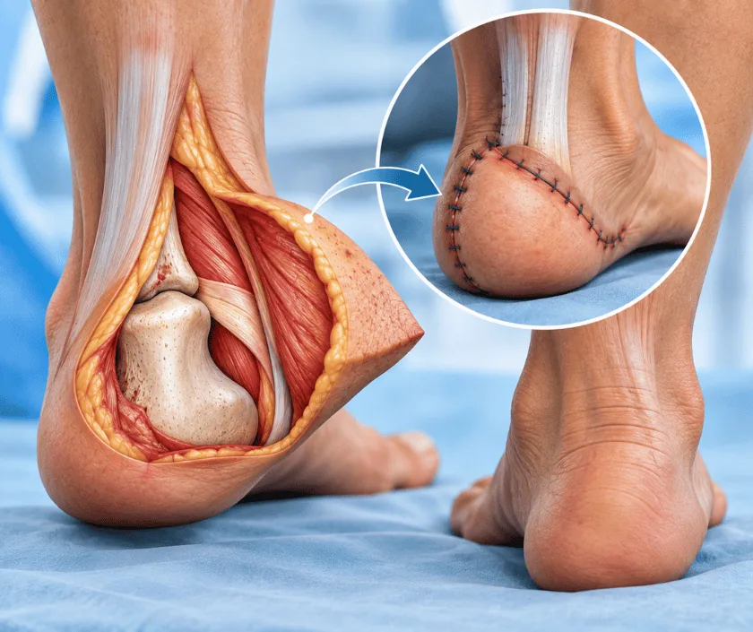
Heel reconstruction replaces skin and padding lost from the back or underside of the heel. Ordinary skin will not survive there for long, because the heel takes body weight with every step. Cover has to be thick enough to cushion, firmly anchored so it does not shear, and durable over years of walking.
Tissue under the heel is unlike skin anywhere else. Fat sits inside tough fibrous chambers that stop it sliding away, and this arrangement absorbs load while holding its shape. When injury, pressure or infection destroys that pad, the heel bone can be left with very little between it and the ground. Simple closure or a graft will not last, because whatever is placed there has to survive repeated loading.
Reconstruction therefore aims at durability rather than appearance. Options include local flaps that bring nearby tissue across, flaps moved on named vessels from the calf, and free tissue transfer where vessels are joined under a microscope. Where sensation can be preserved or restored, that is preferred, since a heel which feels pressure protects itself.
The rest of the picture matters just as much. Circulation in the leg is checked, blood sugars are reviewed and the bone underneath is assessed for infection. Footwear and offloading are planned from the start, because even a good flap can fail if pressure is not managed afterwards.
Heel reconstruction suits people whose circulation supports healing and who can keep weight off the area while it settles.
The wound, the circulation and the bone underneath are all examined. Scans of the vessels and of the heel bone are arranged when the picture is not clear from examination alone.
Dead tissue and any infected bone are removed under anaesthetic. Samples go for testing so antibiotics can be chosen accurately rather than guessed.
A local flap, a flap from the calf on named vessels or a free flap is selected. Durability under load and preserved sensation carry more weight than appearance here.
Tissue is moved into the defect and secured so it cannot shear sideways once walking eventually resumes. The donor area is closed directly or grafted.
A boot, cast or wheelchair regime is arranged before you go home, since keeping pressure off the heel is what protects the reconstruction.
The foot is elevated and kept entirely off the ground. Flap colour is checked frequently and pain relief is given regularly so rest is possible.
Dressings are reviewed and the donor site is checked. Transfers into a chair are taught, and going home is usual once the flap is stable.
Loading begins carefully in a protective boot if the wound has healed. Swelling continues to vary through the day and elevation still helps.
The flap toughens with gradual use. Footwear is reviewed, insoles may be prescribed, and any thinning procedure is only considered at this stage.
A good reconstruction gives healed, durable cover that tolerates walking in suitable footwear. It will not reproduce the natural heel pad exactly, so cushioning feels different and insoles are often needed. Sensation is usually reduced, which means daily checks become part of life. Breakdown can happen years later if pressure is not managed, so long term care matters.
The heel is a demanding site, and the risks reflect that. Discuss each of these before deciding.
Protecting the reconstruction is a long term habit rather than a few weeks of caution, especially where sensation is reduced.
Grafts are thin and will not tolerate walking load. They commonly break down, which is why flap cover is preferred here.
Reconstructed tissue is durable but never identical to the natural pad. Footwear and pressure care remain necessary long term.
Loss of sensation is exactly why some ulcers grow unnoticed. Absence of pain often signals a bigger problem, not a smaller one.
Early loading is what causes breakdown. Strength develops gradually and only after the wound has healed properly.
Heel wounds are treated as a walking problem rather than only a skin problem, so circulation, bone and footwear are considered together. Elegance Clinic explains what the heel will realistically manage afterwards.

The estimate depends on the type of flap chosen, whether infected bone needs clearing, the number of theatre visits, hospital stay and the offloading equipment required afterwards. Cases involving diabetes or circulation problems often need extra investigations, which are listed separately.
A written estimate is prepared after assessment. If insurance applies, the office will explain which documents are usually requested and where approval may take time.
These are the questions that come up most often in consultation. If yours is not here, send it on WhatsApp and the team will reply, usually the same day.
Ask your question →Body weight passes through the heel at every step, and walking also pushes tissue sideways. The natural pad is a specialised cushion that no transferred tissue reproduces exactly, so cover must be chosen for durability rather than appearance.
A written estimate is given after assessment. The figure reflects the flap chosen, whether bone needs clearing, the length of stay and the offloading equipment needed. Investigations for circulation and diabetes are itemised so nothing comes as a surprise.
Loading usually begins carefully at around six weeks in a protective boot, provided the wound has healed. Full walking takes longer. Rushing this stage is the most common reason a reconstruction breaks down, so the schedule is worth respecting.
Sensation is usually reduced and rarely returns fully. That matters practically, because a heel which cannot feel pressure may be injured without you noticing. Daily inspection and prescribed footwear become part of ordinary routine afterwards.
Often yes, though sugar control and circulation are assessed first. Poor control slows healing and increases the risk of infection. A physician is involved throughout, and surgery is timed for when the general picture supports healing.
It can, particularly if pressure is not managed or sensation is poor. Suitable footwear, insoles and regular checks reduce that chance considerably. Any new redness or blister should be reviewed early rather than watched at home.
Bring previous wound photographs, imaging, discharge summaries, diabetes records and a list of medicines. Wearing the shoes you use daily is helpful, since footwear often forms part of both the cause and the solution.
Each technique below has its own page explaining how it works and when it is chosen.
Seeing patients from across the city and beyond: read about the practice on the plastic surgeon in Surat page, or check what a written estimate covers on the costs and insurance page.
