When a segment of the shin bone is lost to injury or infection, the leg cannot carry weight until that gap is rebuilt. Several techniques exist, and the right one depends on the size of the gap and the state of the soft tissue.
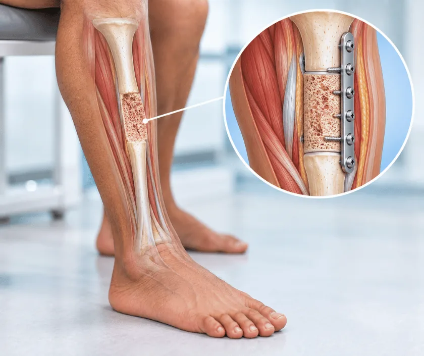
Tibial defect reconstruction rebuilds a missing segment of the shin bone. Small gaps may fill with bone graft, while larger ones need bone transport, a staged membrane technique or a bone flap carried on its own blood vessels. Healthy soft tissue cover has to be in place first, because bone will not heal while exposed.
The tibia is the main weight bearing bone of the lower leg. Losing a section of it, whether at the time of an accident or when infected bone has to be removed, leaves the leg unable to support the body. Rebuilding that section is possible, but the method has to match the size of the gap and the quality of the tissue around it.
Short gaps often heal with bone graft taken from the pelvis, held in place by a plate, nail or frame. As the gap grows longer, graft alone becomes unreliable, so other approaches are used. Bone transport slides a segment of the patient own bone slowly across the gap. A staged membrane technique first places a spacer, then fills the space with graft at a second operation. Where blood supply is poor, a bone flap carried on its own vessels can be transferred from the fibula.
Whatever the method, two conditions come first. Infection must be cleared, and the bone must be covered by healthy, well supplied soft tissue. Skipping either step usually leads to failure.
Bone reconstruction suits people whose infection is controlled, whose soft tissue is healthy and who can commit to a long treatment plan.
Imaging shows the length and position of the gap. Blood tests and samples help judge whether infection is still active, and the soft tissue envelope is examined carefully.
Dead and infected bone is removed until healthy bleeding bone is reached. This may make the gap larger, but grafting into infected bone is rarely successful.
Any exposed area is covered with a flap so the bone sits in a well supplied envelope. Bone reconstruction is generally deferred until this has healed.
Graft, bone transport, a staged membrane technique or a bone flap is selected according to the size of the gap, the quality of the tissue and your general health.
The chosen method is carried out and the leg is held with a frame, nail or plate. Regular imaging then tracks how the new bone is forming.
The leg is elevated and rested. Pain relief and antibiotics are given, and pin site or wound care is taught before you become more mobile.
Dressings and pin sites are reviewed. Walking with crutches usually begins under supervision, and going home is common once care is understood.
Imaging checks how the bone is responding. Weight bearing is adjusted, and physiotherapy works on knee and ankle movement alongside.
New bone continues to form and strengthen. The frame or fixation stays until imaging shows the bone can take load, which often takes many months.
Bone reconstruction can restore a leg that carries weight, but it asks for patience. Treatment often runs for many months and further procedures are common. Some difference in length or alignment may remain, and stiffness in nearby joints is usual. Recovery can vary a great deal, so progress is judged on imaging and function rather than on a fixed calendar.
Rebuilding bone is a long process, and problems along the way are not unusual. Knowing them helps you plan.
Most of this treatment happens at home between reviews, so daily care has a direct effect on the outcome.
Only short gaps behave that way. Longer ones need transport, a staged technique or a bone flap to heal reliably.
Exposed bone becomes infected instead of healing. Soft tissue cover comes first for that reason.
Comfort is not proof of strength. Imaging decides when the new bone can safely take load without support.
Nutrition helps, yet mechanics, blood supply and freedom from infection matter far more than any supplement.
Bone gaps are planned alongside the soft tissue cover they depend on, so the sequence makes sense from the outset. Elegance Clinic explains the expected timeline before treatment starts.

The estimate reflects the method chosen, the implants or frame required, the number of operations and the length of stay. Bone transport and staged techniques involve more reviews and often more imaging, and those items are listed so the picture is complete.
A written estimate is prepared after assessment. Where the injury falls under accident cover or an employer scheme, the office will explain what documents insurers usually ask for.
These are the questions that come up most often in consultation. If yours is not here, send it on WhatsApp and the team will reply, usually the same day.
Ask your question →Months rather than weeks, and longer gaps take longer still. New bone forms slowly and has to strengthen before it takes full weight. Imaging at each review guides when the frame or fixation can safely come off.
Flap cover after an open fracture sits in the band Rs 1.8L to Rs 4.5L, and the figure is case based. The estimate is prepared in writing after assessment, covering the operations planned, implants or frame and hospital stay.
Bone left exposed dries and becomes infected instead of healing. A flap brings circulation and seals the area, giving grafted or transported bone a healthy environment. Reversing the order usually leads to failure.
Some difference in length can remain, though techniques such as bone transport are designed to preserve length. Where a difference persists, a shoe raise usually compensates well. This is discussed before treatment rather than afterwards.
Not directly. Infected and dead bone must be removed first and the infection treated, which often makes the gap bigger. Once samples and blood tests suggest it is settled, reconstruction can proceed more safely.
It is bulky and takes adjustment rather than being constantly painful. Pin sites can be sore, especially early on, and clothing needs modifying. Most people adapt within a few weeks with support and clear instructions.
Imaging and previous operation notes are reviewed, the leg is examined and the soft tissue is assessed. Options are explained with their timelines and limits. Blood tests are usually arranged, and a written estimate follows.
Each technique below has its own page explaining how it works and when it is chosen.
Seeing patients from across the city and beyond: read about the practice on the plastic surgeon in Surat page, or check what a written estimate covers on the costs and insurance page.
