One of the two bones in the lower leg can be borrowed, with its blood vessels, to rebuild a missing section of jaw. Because the transferred bone stays alive, it heals, resists infection and can later hold dental implants.
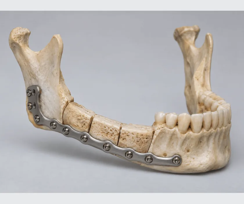
A free fibula flap uses the slender outer bone of the lower leg to rebuild the jaw. Bone, its blood vessels and often a paddle of skin are moved to the face, and the vessels are joined under a microscope so the graft stays alive. Because the fibula carries little body weight, the leg usually functions well afterwards, though it needs its own recovery.
Two bones run between the knee and the ankle. The thick inner one, the tibia, carries the weight of the body. The slender outer one, the fibula, mainly serves as an anchor for muscles and as part of the ankle joint. Surgeons can remove a long central section of the fibula while leaving the ends that matter, which is why it has become the workhorse for rebuilding the jaw.
During surgery one team works on the face while another raises the bone in the leg. The fibula is cut into segments and shaped so that its straight length recreates the natural curve of the jaw. A plate holds the segments in position. Blood vessels from the leg are then joined to vessels in the neck under a microscope, and within minutes the bone is alive in its new home.
The leg is closed with a graft or directly, depending on how much skin was taken. Standing and walking with support usually begin within a few days, and full comfort in the ankle and foot takes considerably longer.
Suitability depends as much on the leg as on the jaw. The circulation of the lower limb is checked carefully before the operation is offered.
Pulses are examined and imaging of the leg vessels is usually arranged, because the fibula can only be taken safely when the remaining arteries supply the foot well. The opposite leg is assessed too.
Scans of the jaw and the leg are used to plan where the bone will be cut. Guides or a printed model help recreate the jaw curve, which improves the bite and shortens theatre time.
A team lifts the fibula with its artery, vein and, when needed, a paddle of calf skin. Ends of the bone near the knee and ankle are deliberately left in place to protect stability.
Wedges are removed from the bone so it bends into the jaw curve, and a plate fixes the segments to the remaining jaw. Position is checked against the bite before the vessels are joined.
Artery and vein are joined to neck vessels under a microscope, and the flap is watched closely afterwards. The leg is closed, sometimes with a skin graft, and a splint supports the ankle initially.
The flap is checked frequently because early problems can often be rescued. The leg is kept elevated in a splint, and feeding usually runs through a fine tube.
Standing and walking with a frame or crutches typically begin, guided by the physiotherapist. Neck drains come out, swallowing trials start and discharge is often possible.
Most people walk without support by now, though the ankle can still feel stiff and the leg swells by evening. Soft diet continues and jaw healing is reviewed.
Bone has usually united and the leg feels much closer to normal. Dental implants, when suitable, are generally discussed from around this point.
Jaw shape and bite alignment are usually restored well, and most people return to eating a normal range of soft and moderate foods. The calf carries a long scar, the ankle may feel stiff and the leg often swells at the end of the day for many months. Some numbness on the outer foot is common. Dental restoration is staged and not always achievable. Outcomes can vary widely between patients.
Two surgical sites mean two sets of risks, and the donor leg deserves as much attention in the discussion as the jaw.
Two areas need care at once, and the leg is the part people tend to neglect after they get home.
The fibula carries little body weight, and the ends nearest the knee and ankle are left in place. Most people walk unaided within weeks, though the leg needs its own recovery.
The calf wound, ankle stiffness and evening swelling can trouble people for months. Physiotherapy, elevation and compression genuinely change how the leg feels a year later.
Dental restoration is staged. Bone must unite and settle first, gum tissue must be prepared, and radiotherapy may delay matters further. Planning starts early but placement comes later.
A graft without a blood supply struggles across long gaps and in irradiated tissue. Living bone continues to heal and remodel, which is exactly why this flap is chosen.
Microsurgical jaw reconstruction at Elegance Clinic in Surat is planned with the donor leg examined and discussed in as much detail as the jaw itself, so patients know what both sites will ask of them.

Free flap reconstruction of the jaw usually falls in a band of Rs 1.8L to Rs 4.5L. The position within that band reflects the length of bone needed, whether a skin paddle is included, operating time, intensive monitoring afterwards and the plates or custom guides used. Dental implants and prosthetic teeth are quoted separately as a later stage. A written estimate is given before admission and insurance is assessed case by case.
These are the questions that come up most often in consultation. If yours is not here, send it on WhatsApp and the team will reply, usually the same day.
Ask your question →This surgery usually falls in a band of Rs 1.8L to Rs 4.5L. Bone length, skin paddle, operating time, monitoring afterwards and the plates used all influence where a case lands. Dental implants are quoted separately as a later stage.
Cover is assessed case by case. Reconstruction after tumour, injury or bone infection is medically necessary and generally approved with biopsy reports, scans and the surgical plan. Implants and prosthetic teeth are often excluded, so check that clause before admission.
The fibula carries little body weight, and the segments nearest the knee and ankle are left untouched. Leg circulation is checked with imaging beforehand, because the operation is only offered when the remaining vessels supply the foot well.
Most people stand and walk with a frame or crutches within a few days, guided by a physiotherapist. Walking unaided usually follows over the next few weeks. Ankle stiffness and evening swelling can continue for several months afterwards.
Many people describe a long scar, some numbness along the outer lower leg and foot, and swelling that appears by the end of a busy day. Strength and balance usually return well with regular exercises and compression.
Implants are considered only after the bone has united and settled, which normally takes several months. Radiotherapy, gum condition and bone height all affect the decision. A denture is a reasonable alternative when implants are not advisable.
Expect an examination of leg pulses, imaging of the leg vessels, scans of the jaw, blood tests and a physician review. Both legs are assessed. Bring previous operation notes, biopsy reports and a list of your medicines.
Each technique below has its own page explaining how it works and when it is chosen.
Seeing patients from across the city and beyond: read about the practice on the plastic surgeon in Surat page, or check what a written estimate covers on the costs and insurance page.
