Taking out a jaw tumour safely usually means removing bone along with it, which leaves a gap that will not close on its own. Jaw tumour reconstruction fills that gap so the bite, the chin and the ability to chew are restored.
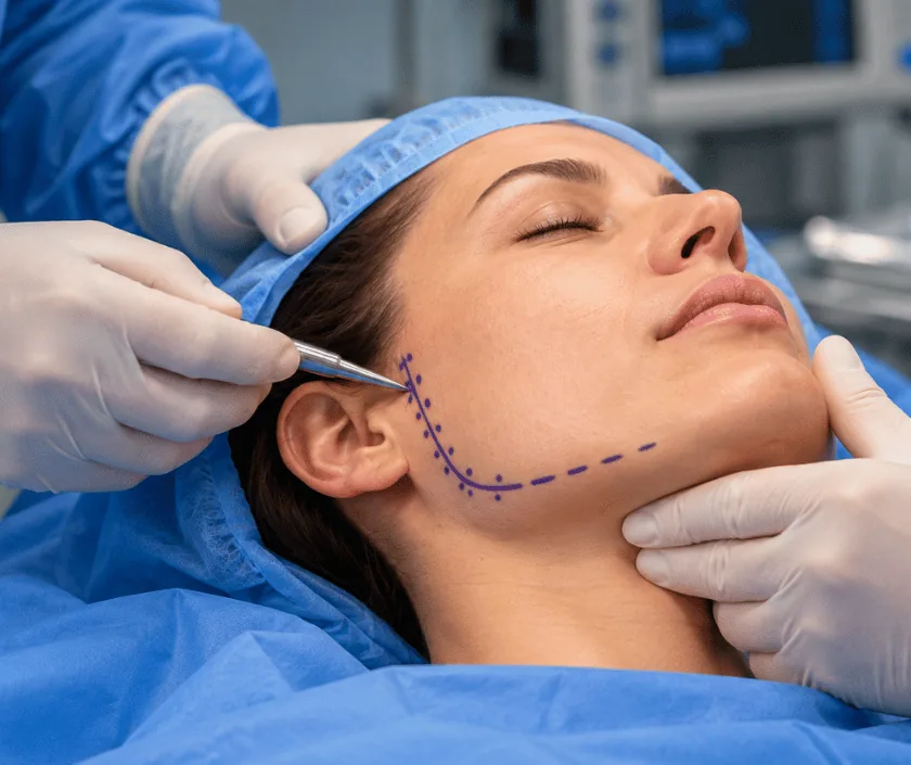
Jaw tumour reconstruction rebuilds the mandible or maxilla after a growth has been removed. Tumours of the jaw may be benign or malignant, and either kind can require a segment of bone to be taken. Living bone with its own blood supply is usually transferred to fill the gap, restoring the jaw curve, the bite and the outline of the face.
Growths in the jaw range widely. Some are cysts that can be removed simply, some are benign tumours that invade bone locally, and some are cancers that need wide clearance along with treatment of the neck. What they share is that once a full thickness section of jaw is taken, the two remaining ends drift, the bite goes out of line and the face changes shape. That is the problem reconstruction exists to solve.
Planning starts with a biopsy and a scan, so that the type of tumour and its extent are both known before any decision. The removal is then designed with a margin appropriate to the diagnosis, since a benign lesion and a cancer call for different clearances. Where the tumour reaches the lining of the mouth or the skin, those layers are removed too and have to be replaced.
Reconstruction follows in the same sitting. Living bone, most often from the lower leg, is shaped to the original curve and fixed with a plate, and soft tissue is brought in with it when lining or skin is missing.
Nearly everyone losing a segment of jaw benefits from reconstruction, though the method is matched to the diagnosis, the defect and the fitness of the patient.
Biopsy establishes the type of tumour, and scans show how far it extends into bone and soft tissue. For cancers the neck is assessed as well, since treatment there is planned at the same time.
Margin is chosen according to the diagnosis. Cutting guides or a printed model help place the bone cuts precisely, which makes the reconstruction fit better and shortens theatre time.
The jaw segment is taken out with the planned margin, along with any teeth and lining involved. Samples are checked during surgery where the facility allows.
Living bone is raised with its vessels, shaped to the jaw curve and fixed with a plate. The vessels are joined to neck vessels under a microscope so the bone survives.
The reconstruction is monitored closely in the early days. Speech, swallowing and mouth opening therapy begin as healing allows, and dental planning follows months later.
Monitoring of the reconstruction dominates. The face and neck are swollen, feeding is often through a fine tube and a drain is usually present.
Drains and tubes are removed, swallowing trials start and mouth care is taught. Most people are ready to go home towards the end of this stretch.
Soft diet is generally established and mouth opening exercises are under way. If radiotherapy is planned, it commonly begins once wounds have healed.
Bone has usually united and swelling has settled. Dental rehabilitation and any contour refinement are considered from this stage onwards.
Shape and bite alignment are usually restored well, and most people return to a reasonable range of foods. Numbness of the lip or chin is common where the nerve was involved, and scars on the neck and donor site remain visible. Radiotherapy tightens tissues and can limit mouth opening. Dental restoration takes months and is not always possible. Outcomes can vary a great deal, so progress is reviewed honestly at each visit.
Reconstruction and removal are one operation with two sets of risks, and both are explained before consent.
Consistent mouth care, good nutrition and attending review visits protect both the reconstruction and your longer term health.
Some benign tumours invade bone locally and return when treated too lightly. The extent of removal follows the behaviour of the tumour, not simply its label.
Rebuilding at the same sitting keeps the jaw aligned and lets radiotherapy start on time. Delayed reconstruction is harder, because the ends of the bone drift and scar.
Plates alone can work across short defects at the back of the jaw, yet in the front or in irradiated tissue they tend to loosen, break or become exposed over time.
Follow up scans, therapy, dental work and checks for recurrence continue for years. Wound healing is an early milestone rather than the end of care.
Jaw tumour care at Elegance Clinic in Surat brings removal and reconstruction into one plan, with scans, models and the dental endpoint all discussed before the date of surgery is fixed.

Free flap reconstruction of the jaw usually falls in a band of Rs 1.8L to Rs 4.5L. The exact figure reflects the length of bone removed, whether soft tissue is transferred with it, operating time, monitoring afterwards and the plates or planning guides used. Dental implants and prosthetic teeth are quoted separately as a later stage. A written estimate is given before admission, and insurance is assessed case by case.
These are the questions that come up most often in consultation. If yours is not here, send it on WhatsApp and the team will reply, usually the same day.
Ask your question →Free flap reconstruction of the jaw usually falls in a band of Rs 1.8L to Rs 4.5L. Length of bone removed, operating time, monitoring and the plates used all influence the figure. Dental implants and teeth are quoted separately as a later stage.
Cover is assessed case by case. Removal and reconstruction of a jaw tumour are medically necessary, so approval is usually granted with the biopsy report, scans and surgical plan. Dental implants are commonly excluded, and policy sub limits still apply.
This is established surgery, though it is long and needs good heart and lung fitness. Flap failure, infection and donor site problems are the principal risks. Blood tests, scans and a physician review are completed before anaesthesia is agreed.
Around a week is usual when a free flap is used, because the reconstruction needs close monitoring in the early days. Simpler repairs need less. Discharge depends on swallowing, wound healing and how steady you feel on your feet.
Most people speak clearly and eat a reasonable range of foods once healing and therapy are complete. Chewing strength and mouth opening may be somewhat reduced, particularly after radiotherapy, and regular exercises help considerably.
Recurrence is possible with both benign and malignant jaw tumours, which is why surveillance continues for years. Regular examination and scans catch a return while it is small, so every scheduled appointment is worth keeping.
Bring the biopsy report, all scan films and reports, previous operation notes and a list of your medicines. If you are already under an oncology team, bring their letters and any treatment schedule as well.
Each technique below has its own page explaining how it works and when it is chosen.
Seeing patients from across the city and beyond: read about the practice on the plastic surgeon in Surat page, or check what a written estimate covers on the costs and insurance page.
