Lower jaw bone gives the chin its projection, holds the lower teeth and drives every bite you take. Losing a section of it changes speech, eating and appearance together, which is why reconstruction is planned so carefully.
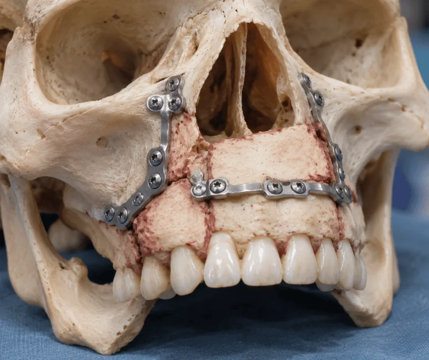
Mandibular reconstruction rebuilds the lower jaw after tumour removal, injury or infection. Because the mandible is a moving bone that carries teeth, the reconstruction must restore both its shape and its ability to work. Options run from a titanium plate alone, through bone grafts, to a free flap that brings living bone with its own blood supply.
Removing a segment of lower jaw leaves a gap that behaves very differently from a gap anywhere else. The jaw is a single curved bone that moves thousands of times a day, so the two remaining ends drift, the bite goes out of line and the chin loses its projection. Speech and chewing suffer immediately, and the change to the profile is usually obvious.
Reconstruction is planned as a ladder of options. For a short defect at the back of the jaw, a strong titanium plate may bridge the gap adequately, particularly in an older patient who wants minimal surgery. Where healthy tissue surrounds a small gap, a bone graft taken from the hip can fill it. For longer defects, for the front of the jaw, and whenever radiotherapy has been or will be given, living bone with its own blood supply is the more dependable answer, and the lower leg is the usual source.
Planning now often uses a three dimensional model, so the new bone can be cut and shaped to match the original curve before the operation even starts.
Choice of technique depends on where the defect sits, how long it is, whether radiotherapy is planned and how fit the patient is for a long operation.
A CT scan of the jaw and often of the donor limb guides the plan. A three dimensional model lets the surgeon plan the cuts and bend the plate before surgery, which shortens the operation.
The affected segment is taken out with a clear margin, and teeth in the segment are removed with it. Nerve position and the future bite are considered while planning the cuts.
Where a free flap is used, bone is raised with its blood vessels and cut into segments that recreate the curve of the jaw. A plate holds the shape while it is fixed in place.
The reconstruction is fixed to the remaining jaw so that the bite lines up. Blood vessels are joined to vessels in the neck under a microscope, which keeps the new bone alive.
The flap is monitored closely in the first days. Once bone has united over the following months, implants or a denture are planned to restore chewing properly.
Close monitoring of the flap dominates these days. Feeding usually runs through a fine tube, the neck is swollen and a drain is often in place.
Drains and tubes are removed as things settle. Swallowing trials begin, mouth care is taught, and most people are ready to go home towards the end of this period.
Bone healing is under way and diet has usually widened to soft food. Any planned radiotherapy is commonly started once wounds have healed.
Bone has generally united and swelling has settled. Dental implants, if they are suitable, are usually considered from around this stage onwards.
Most patients regain a stable jaw, a workable bite and a recognisable facial outline. Chewing improves considerably once teeth are restored, though it may never feel quite as strong as before. Numbness of the lower lip is common and may persist, and scars on the neck and donor limb remain visible. Radiotherapy adds stiffness. Recovery can vary widely, so goals are set individually and reviewed at each stage.
This is major surgery with a donor site as well as a reconstruction site, and both carry risk.
Mouth hygiene and steady nutrition protect the reconstruction during the months that bone takes to unite.
A plate can bridge short gaps at the back of the jaw, but over time plates in the front or in irradiated tissue tend to loosen, break or become exposed.
A hip graft suits small defects with healthy surroundings. Without its own blood supply it struggles across long gaps and after radiotherapy, where living bone performs far better.
Immediate implants are occasionally possible, yet in most cases the bone must unite and settle first. Rushing risks losing the implants and disturbing the reconstruction.
Most people eat, speak and return to ordinary life. Function is usually good, though chewing strength and mouth opening may be somewhat reduced.
Jaw reconstruction at Elegance Clinic in Surat is planned with scans and models before the day of surgery, and dental rehabilitation is discussed at the very first visit rather than left until afterwards.

Free flap reconstruction of the lower jaw usually falls in a band of Rs 1.8L to Rs 4.5L. Where the figure lands within that range depends on the length of the defect, the operating time, intensive monitoring afterwards, plates and any custom planning. Dental implants and teeth are quoted separately, since they come later. Insurance cover is assessed case by case and a written estimate is given before admission.
These are the questions that come up most often in consultation. If yours is not here, send it on WhatsApp and the team will reply, usually the same day.
Ask your question →Free flap reconstruction of the jaw commonly falls in a band of Rs 1.8L to Rs 4.5L. Defect length, operating time, monitoring afterwards and the plates used all move the figure. A written estimate is given before admission, with dental work quoted separately.
Cover is assessed case by case. Reconstruction after tumour, injury or infection is medically necessary and usually approved with the biopsy report, scans and surgical plan. Dental implants are frequently excluded, so read that section of your policy before you proceed.
Jaw reconstruction with a free flap is an established operation, though it is long and demands good heart and lung fitness. Flap failure, infection and problems at the donor site are the main risks. Fitness is assessed with blood tests, scans and a physician review.
Hospital stay is often around a week with close flap monitoring. Soft diet continues for several weeks and bone usually unites over a few months. Donor site physiotherapy and mouth opening exercises carry on through much of that period.
Most people chew comfortably once teeth are restored, though strength may be somewhat less than before. Mouth opening can be reduced, especially after radiotherapy. Regular exercises and a well planned dental result make the largest practical difference.
Often yes, but only after the reconstructed bone has healed and settled, which usually takes several months. Radiotherapy, bone height and gum condition all influence the decision. A denture is a reasonable alternative when implants are not advisable.
Expect a mouth examination, a bite assessment, review of your scans and a discussion of donor site options. Photographs and impressions may be taken. Bring biopsy reports, previous operation notes and a list of your medicines to that visit.
Each technique below has its own page explaining how it works and when it is chosen.
Seeing patients from across the city and beyond: read about the practice on the plastic surgeon in Surat page, or check what a written estimate covers on the costs and insurance page.
