Of the four walls around the eye, the outer one is the thickest, so breaking it takes real force. Fractures here usually arrive as part of a wider cheek and socket injury.
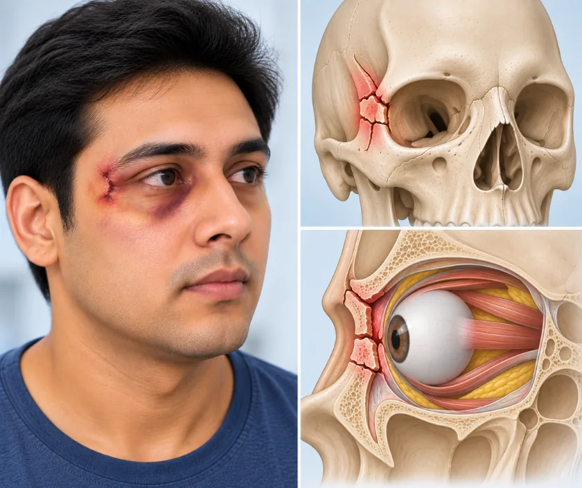
A lateral orbital wall fracture is a break in the strong outer wall of the eye socket, most often part of a larger cheekbone injury. Since the outer corner of the eyelid attaches to this bone, the eye can look tilted or set back once the wall moves. Repair restores the width and the depth of the socket so the eye returns to its proper place.
The outer wall of the eye socket is built for strength. It is formed largely by the cheekbone, which joins the forehead above and the temple behind, and it protects the eye from blows arriving from the side. Because of that thickness, a fracture here signals a forceful injury, typically a road crash, a fall from height or a heavy blow to the side of the face.
Two things follow from the wall moving. Firstly, the socket changes shape, and since the eye rests within that space, it can end up sitting deeper, lower or slightly turned. Secondly, the outer corner of the eyelid loses its anchor, so the eye may look tilted and the outer corner may droop. Bruising, a flattened cheek and numbness over the cheek and upper lip often accompany these changes, and opening the mouth may feel tight when the displaced bone presses on the muscle that works the jaw.
Assessment therefore covers the eye and the cheek together. Vision, eye movement and eye position are checked, and a CT scan shows how far the wall and the surrounding bone have moved.
Repair is advised when the wall has moved enough to change the shape of the socket, the position of the eye or the corner of the eyelid. Undisplaced fractures are often left alone.
Vision, eye movement, eye position, cheek contour and mouth opening are all recorded. The CT scan then shows how far the wall and the cheekbone have moved, and in which direction.
Hidden incisions are used, placed inside the upper eyelid crease, inside the lower lid, inside the mouth or within the hairline, so the face is left without obvious scars.
The cheekbone and the outer wall are lifted back into place and checked at several points at once, since correcting one part and not another leaves the socket the wrong shape.
Small titanium plates and screws hold the position while the bone knits. Where the socket floor or inner wall has also given way, a thin implant is added to span the gap.
The eyelid corner is repositioned if it has been detached, wounds are closed and vision is checked as soon as you wake and again the next day.
Swelling and bruising around the eye and cheek reach their peak. Vision is checked regularly, soft food is easier, and keeping the head raised helps the swelling drain.
Bruising fades and mouth opening improves. Most people manage quiet activity at home, although the cheek stays numb and chewing anything firm is uncomfortable.
Bone has healed enough for normal eating. Eye position and cheek contour are reassessed, and desk work is usually comfortable by this stage.
Final shape becomes clear as the last swelling goes. Numbness of the cheek may still be improving, and any further adjustment is only considered now.
Facial shape usually improves markedly once the bone is back in position, and most people are satisfied that they look like themselves again. Small differences in cheek projection or eye level can remain, especially after a shattering injury. Numbness over the cheek and upper lip often recovers slowly and sometimes not fully. Double vision, when present, generally settles although review over several months is the honest expectation.
This repair is well established, and knowing where it can fall short helps you weigh it up properly beforehand.
The bone needs protecting while it knits, and a few simple habits keep the repair undisturbed.
The cheekbone forms much of the outer wall of the socket. Move it and the socket changes shape, which is why eye position is measured as carefully as facial contour.
Numbness usually comes from a nerve bruised or stretched at the moment of injury. Sensation often returns slowly over months, and it is checked at every review.
Modern repair uses small plates and screws, so the teeth are generally not wired together. Soft food is advised for a while, but talking and drinking carry on normally.
Once bone knits in the wrong position it has to be cut and moved again. Early repair is simpler, more exact and easier on the person going through it.
Cheek and socket injuries are treated at Elegance Clinic in Surat as one problem rather than two, with eye position measured alongside facial contour before and after surgery.

Charges depend on how much of the cheek and socket needs repositioning, the number of plates used, whether an implant is needed for another wall and how long you stay in hospital. Those details come from the scan and the examination, so an estimate is provided in writing afterwards. Repair after an injury is normally covered by health insurance and accident policies, and the team assists with the paperwork.
These are the questions that come up most often in consultation. If yours is not here, send it on WhatsApp and the team will reply, usually the same day.
Ask your question →It depends on how much bone needs repositioning, the number of plates, whether an implant is needed for another wall and the length of stay. A written estimate is prepared after the scan and examination rather than before them.
Injury repair counts as medical treatment, so most health policies and accident cover include it. Emergency records, imaging reports and photographs are usually needed for approval. Waiting periods and room rent limits set out in your policy still apply.
The repair is well established and done under general anaesthesia, with vision checked closely afterwards. Risks worth knowing include lasting numbness, double vision, infection around the plates and a change in lower eyelid position. All are discussed before consent.
Swelling settles over a fortnight and soft food is advised for several weeks. Many people return to light work within three to four weeks. Contact sport waits much longer, and clearance is given at review rather than by a fixed date.
Shape usually improves a great deal once the bone is back in place. Small differences in cheek projection or eye level can remain after a severe injury. Final appearance is judged at around six months, when swelling has fully gone.
No. A fracture that has kept its position, with a level eye and normal cheek contour, is watched. Repair is advised for displaced bone, a tilted or sunken eye, a flat cheek or restricted mouth opening.
Bring scan images and reports, hospital papers from the injury, insurance documents and a list of your medicines. Photographs taken before the injury are genuinely useful, because they show what your own natural symmetry looked like.
Seeing patients from across the city and beyond: read about the practice on the plastic surgeon in Surat page, or check what a written estimate covers on the costs and insurance page.
