The floor of the eye socket is thin enough to give way under a sudden blow, letting soft tissue drop into the sinus below. Double vision, a numb cheek and an eye that sits deeper are the usual clues.
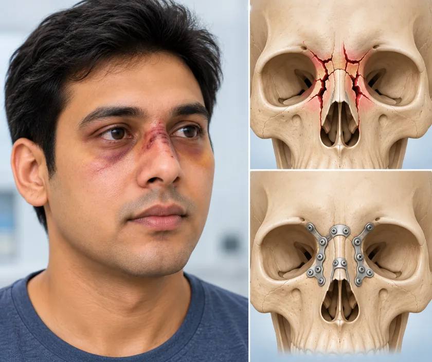
A blowout fracture of the orbital floor is a break in the thin bone under the eyeball, usually after a punch, a ball or a fall. Fat and muscle can slip through the gap into the sinus beneath, which causes double vision, a numb cheek and later an eye that sits deeper than the other. Some settle without surgery, while a trapped muscle needs prompt attention.
Each eye sits in a bony cone with four walls. The floor of that cone is only a fraction of a millimetre thick in places, and directly beneath it lies the air space of the cheek. When a fist, a ball or an elbow strikes the eye, pressure inside the socket rises sharply and the floor gives way, rather like a safety panel. That is where the name blowout comes from, and the eye itself stays in place.
Once the floor has broken, fat and sometimes the muscle that turns the eye upwards can slip through the opening. Double vision follows, usually worst when looking up, and the affected eye may not travel as far as its partner. A nerve running along the floor is often bruised, leaving the cheek, upper lip and upper teeth numb on that side. As swelling settles over the following weeks, the eye may look deeper set than before, because the socket has become larger than the tissue filling it.
Vision, eye movement and sensation are all tested, and a CT scan measures the size of the defect. Together those findings decide whether the injury can be watched or needs repair.
Many small blowout fractures settle with observation as swelling goes down. Repair is offered when the eye is likely to end up sunken or double vision is not improving.
Vision, eye movement, pupil reactions and sensation over the cheek are recorded first. A CT scan then shows the size of the gap in the floor and what has dropped through it.
Urgent surgery is arranged when tissue is trapped. Otherwise repair is usually planned within about two weeks, once swelling has fallen far enough for the eye position to be judged.
The floor is reached through an incision inside the lower eyelid or in a natural crease, so the scar is hidden. The socket is opened carefully with the eye protected throughout.
Fat and muscle are lifted gently back into the socket, and a thin implant is shaped to span the gap. Once in place, the eye is moved by hand to confirm nothing is still caught.
The wound is closed and vision is checked as soon as you wake and again over the next day. Any sudden pain or visual change afterwards is treated as urgent.
Bruising and swelling of the eyelids are at their worst and vision is checked repeatedly. Sleeping propped up helps, and nose blowing is forbidden while the sinus below is healing.
Swelling drops and stitches, if used, come out or dissolve. Double vision usually improves in stages, and screen work is manageable for many people by the end of this period.
Eye movement is reassessed formally. Most people are back at work, while contact sport, swimming and anything risking a knock to the eye are still avoided.
Eye position and any remaining double vision are judged final around now. Numbness of the cheek may still be improving, and further options are discussed only at this stage.
Most people regain comfortable single vision when looking straight ahead and reading, which is what matters day to day. Slight double vision at the extremes of gaze can remain, and it is usually tolerable. Eye position improves although a small difference between the two sides may persist. Numbness of the cheek and upper lip recovers slowly and sometimes incompletely, so it is fair to expect months rather than weeks of change.
Working close to the eye demands caution, and the risks below are discussed openly before consent is taken.
Protecting the eye and keeping pressure out of the socket are the two things that matter most in the early weeks.
Bruising says nothing about the bone underneath. Numbness of the cheek, double vision or trouble looking up are the signs that point towards a fracture and call for a scan.
Despite the name, the eye stays where it belongs. It is the thin floor beneath that gives way, which protects the eyeball from taking the full force.
Plenty of double vision comes from swelling and bruising and clears as those settle. Surgery is considered when it persists, when tissue is trapped or when the eye is sinking.
Late correction is harder because scar tissue holds everything in the wrong place. A trapped muscle in particular is a same day problem, not something to leave for weeks.
Eye socket injuries at Elegance Clinic in Surat are assessed with vision and eye movement recorded from the start, and repair is planned alongside an eye specialist rather than in isolation.

Cost here follows the size of the defect, the type of implant used, whether surgery is urgent or planned and how long you stay in hospital. Those points are settled only after eye assessment and a scan, so the estimate is prepared in writing at that stage. Repair after injury is medical treatment, so health insurance and accident policies normally apply, and the team will help with the documents.
These are the questions that come up most often in consultation. If yours is not here, send it on WhatsApp and the team will reply, usually the same day.
Ask your question →It varies with the size of the gap in the floor, the implant chosen, whether surgery is urgent and the length of stay. A written estimate is prepared after eye assessment and a scan, rather than a figure being quoted in advance.
Usually yes, because this is treatment for an injury and not cosmetic work. Emergency records, the scan report and photographs are generally required for approval. Room rent limits and waiting periods written into your policy continue to apply.
The repair is well established and done under general anaesthesia, with vision checked before you leave the operating area and again through the next day. Loss of vision is rare but real, which is exactly why monitoring is so frequent.
Much of it comes from swelling and improves over two to three weeks. What remains is reassessed at six weeks and again at six months. Recovery can vary, and a small amount at the extremes of gaze may stay.
Restoring the volume of the socket is the main aim, so most eyes end up close to level with the other side. A slight difference can remain after a large fracture, and it is judged honestly once swelling has fully gone.
No. Small breaks with good eye movement are watched and often settle. Repair is advised for trapped tissue, for double vision that is not improving, or for a large defect likely to leave the eye sunken.
Bring the scan images and reports, any emergency papers, spectacles you use, insurance documents and your medicine list. Mention smoking, diabetes or blood thinning tablets. Note when the double vision is worst, as that detail guides the plan.
Seeing patients from across the city and beyond: read about the practice on the plastic surgeon in Surat page, or check what a written estimate covers on the costs and insurance page.
