Some accidents break the forehead, the cheekbones, the upper jaw and the lower jaw in a single event. With no undamaged landmark left to build against, the order in which each part is repaired becomes the heart of the plan.
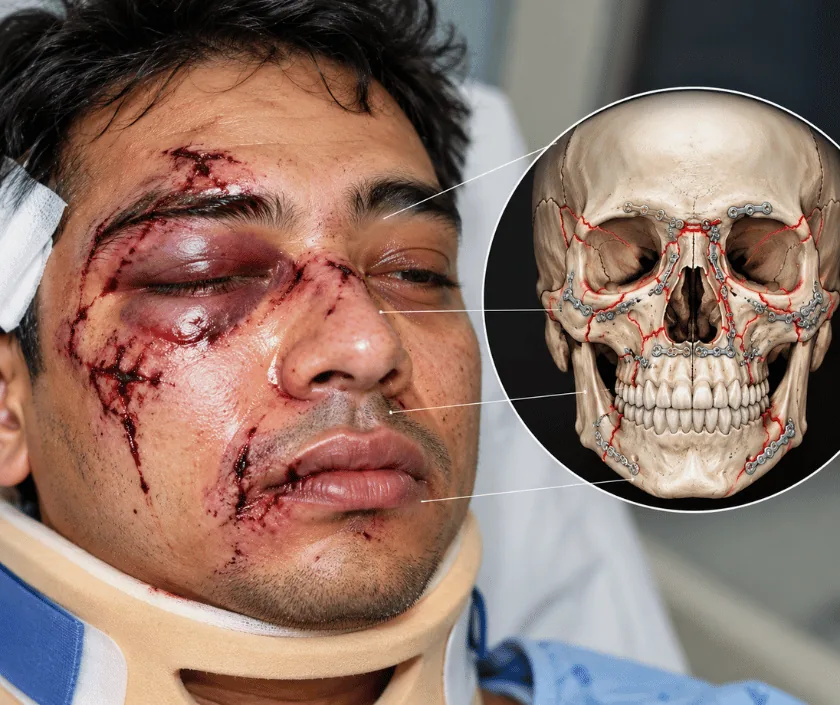
Panfacial fracture management is the treatment of injuries that break the upper, middle and lower face at the same time. Since every usual reference point is broken, each area has to be rebuilt against one that has already been fixed. Sequencing, restoring the bite and protecting the airway matter as much as the individual repairs themselves.
Facial repair normally works outwards from something intact. A cheekbone is set against the eye socket rim, the upper jaw against the bite, the bite against a sound lower jaw. In a panfacial injury none of those references can be trusted, because each of them is broken. The face has lost its scaffolding in every direction, and simply plating each fracture where it lies would lock in a face that is too wide, too flat or too long.
Planning therefore begins with a decision about sequence. Many surgeons start with the lower jaw, restore its width and shape, then use the bite to position the upper jaw, and finally rebuild the outer frame of the face at the cheekbones and the forehead. Others work from the stable skull downwards. Either way, each step is checked before the next is committed, and the scan is used throughout as the map.
Around all of this sit the practical questions. Can the patient breathe safely during and after surgery? Are there head, chest or limb injuries that come first? How much can be achieved in one sitting before the swelling and blood loss make it wiser to stop and continue later?
Everyone with multiple displaced facial fractures needs a planned sequence. What varies is how much is done at one sitting and how soon it begins.
Breathing is protected first, bleeding is controlled and life threatening injuries are treated. A temporary breathing tube in the neck is sometimes needed so the bite can be handled freely during surgery.
A computed tomography scan shows every fracture in the upper, middle and lower face. The plan then sets out which area will be repaired first and what each following step will be measured against.
The first area repaired becomes the reference for everything else, commonly the lower jaw or the stable bone of the skull and outer eye socket rim. Its width and projection are restored carefully.
The teeth are brought into their earlier relationship and held, which positions the upper jaw. Pillars of the midface and the cheekbones are then fixed to reset facial height and width.
If swelling, blood loss or fatigue make continuing unwise, the remaining work is planned for a second sitting. Reviews follow the bite, jaw opening, eye position and facial contour over months.
Care centres on breathing, swelling, pain and nutrition, often in intensive care. The face is heavily swollen and feeding is given in liquid form.
Swelling starts to fall and stitches are removed. Speech and swallowing are reassessed, jaw exercises begin gently and a soft diet is introduced when it is safe.
Fractures have united and any guiding elastics come off. Jaw opening exercises continue in earnest and diet advances step by step.
Facial shape, bite and jaw movement are reviewed properly. Refining surgery, dental replacement or scar treatment may be planned once everything has settled.
Recovery after a panfacial injury can vary a great deal, and progress is measured in months. Careful sequencing gives many patients a working bite, reasonable jaw opening and a face close to its earlier proportions. Some flattening, asymmetry, lasting numbness or a bite needing dental adjustment often remains. Further procedures are commonly part of the plan, and treatment continues after the bone has healed.
Both the injury and its treatment carry substantial risks, and these are set out for the family before consent.
Much of the final result is decided at home over the months after discharge rather than in theatre.
Without a sound reference, that locks in a face that is too wide or too flat. Sequence is what makes the individual repairs add up correctly.
Blood loss, swelling and time under anaesthesia all have limits. Stopping and completing the work at a second sitting is often the safer and more accurate choice.
It is usually a temporary measure that keeps the airway safe and frees the surgeon to set the bite. Most such tubes are removed within a few weeks.
Jaw exercises, dental work, scar care and sometimes refining surgery continue for months. That later phase decides much of the day to day result.
Complex facial injuries at Elegance Clinic in Surat are treated as one continuous plan, from the emergency phase through sequenced reconstruction to later refinement, with the same team following the patient throughout.

Cost here follows the whole injury rather than any single procedure, and it moves with intensive care, the number of areas fixed, hardware, theatre time, staged operations and length of stay. Jaw fracture fixation gives the nearest reference band and is shown below as a guide to the surgical component. A written estimate is prepared once the patient is stable and the sequence is planned, and mediclaim usually applies.
These are the questions that come up most often in consultation. If yours is not here, send it on WhatsApp and the team will reply, usually the same day.
Ask your question →The surgical component usually falls in a band of Rs 55,000 to Rs 1.2L, while the total depends on intensive care, imaging, hardware, staged operations and length of stay. A written estimate is shared with the family once the sequence of repair is planned.
Accident related facial reconstruction is covered by most mediclaim policies, since it is reconstructive rather than cosmetic. Claims need accident records, scan reports and operative notes. Room rent limits, waiting periods and dental exclusions in the policy still apply.
It is planned around what the patient can tolerate, with the airway protected and other injuries treated first. Bleeding, infection, numbness and limited jaw opening are the main risks. Staging the work is often chosen precisely to keep the operation within safe limits.
Hospital stay is often a week or more. Swelling settles over several weeks and bone unites by about six weeks, while jaw exercises and dental work continue for months. Returning to work usually takes two to three months.
Careful sequencing gives many patients a working bite and a face close to its earlier proportions. Some asymmetry, flattening or lasting numbness often remains, and refining procedures are commonly planned once healing is complete.
Swelling, blood loss and time under anaesthesia limit what can be done safely in one sitting. Splitting the work protects the patient and often improves accuracy, since the second stage is planned on a settled face.
Usually within the first one to two weeks, once breathing, bleeding and any head injury are controlled. Earlier is better for accuracy, though patient safety decides the timing rather than the calendar.
Seeing patients from across the city and beyond: read about the practice on the plastic surgeon in Surat page, or check what a written estimate covers on the costs and insurance page.
