An ear torn partly or completely away is one of the few facial injuries where hours genuinely count. Bringing the detached part with you gives the surgical team its best chance of using it, whether by reattachment or as tissue for repair.
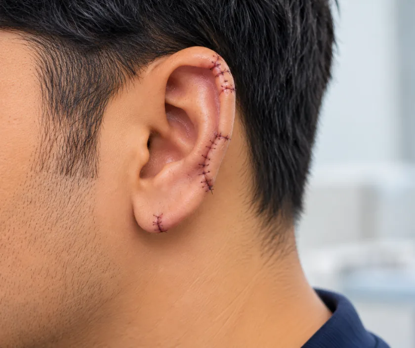
Ear avulsion repair treats an ear that has been torn partly or completely off, usually in a road accident, a bite or a pulled earring. Time matters, because a detached ear may be replantable if it reaches a surgical team quickly. Where replantation is not possible, the ear is rebuilt in stages using cartilage and skin cover.
The outer ear is a delicate frame of cartilage under thin skin, held on by a blood supply that is generous for its size. That generosity helps, because a torn segment attached by only a slim bridge of skin will often survive if it is repositioned early and handled gently. When the ear comes away completely, the picture changes and the clock starts.
A detached ear that reaches a surgical team quickly may be reattached, either by joining the tiny vessels under magnification or, in selected cases, by burying the cartilage under nearby skin so that it can be raised again later. Neither is possible in every injury, since crushed or heavily contaminated tissue cannot be used. What the team can offer depends greatly on how the part was carried and on how much time has passed.
Where the ear cannot be saved, reconstruction is planned in stages once the wounds have healed. Cartilage, usually taken from the rib, is carved into a framework and placed under skin, with later procedures to lift the ear away from the head. The aim is a natural outline that supports spectacles and matches the other side reasonably closely.
What is possible depends on the injury, the time that has passed and the condition of the detached part. Decisions are often made quickly, with reconstruction planned later if reattachment is not an option.
Other injuries are checked first, then the ear and the detached part are examined. The wound is washed, tetanus protection is reviewed, and photographs are taken for the record.
The team judges whether the part is clean enough and whether vessels can be joined. Time since the injury, the crush pattern and how the part was carried all influence that decision.
Where possible the segment is reattached, with fine vessels joined under magnification in selected cases. Partial tears are repaired in layers, so that cartilage is aligned and covered by skin.
If the ear cannot be reattached, the wound is closed with local skin or a graft so that the area heals. Cartilage is preserved where it is clean and usable.
Once healing is complete, an ear framework can be carved from rib cartilage and placed under skin, with further stages to lift and shape the ear.
The ear is protected from pressure and watched closely for colour and warmth. Pain relief and antibiotics are given, and sleeping on that side is avoided.
Stitches are usually removed during this period. Swelling settles, dressings are simplified, and the shape of the repair becomes easier to judge.
The repair is stable though still tender. Spectacles and helmets can often be used again around this stage, guided by your team.
Scars soften and the outline settles. Further stages of reconstruction, or refinements to a repaired rim, are planned from this point onward.
A repaired ear usually keeps a recognisable outline, with scars along the rim or behind the ear. Reattachment can succeed, though it is not possible in every injury, and the result may include some loss of skin or a change in colour. Reconstructed ears look convincing at conversational distance yet differ close up in fine detail. Sensation is often reduced. Several stages are usual, and further refinement is sometimes needed once tissues have settled.
This is delicate tissue with a fine blood supply, so the risks deserve a plain description before any procedure.
A repaired ear needs protection more than attention, and a few rules make a large difference.
Reattachment is possible in selected injuries, and delay removes the option. Bringing the part with the patient keeps that choice open.
Cartilage has no blood supply of its own and depends on the skin covering it. Simply stitching a detached ear back rarely works, which is why the approach is different.
Once the edges have healed over with skin, they will not join. Repair means freshening those edges and closing them in layers.
A rebuilt framework needs healthy, settled skin cover. Reconstruction is planned once the wounds have healed, often several months later.
Ear injuries at Elegance Clinic in Surat are treated as urgent, with assessment of the detached part, secure cartilage cover and a clear plan for any later reconstruction.

Repair of a torn or detached ear is emergency and reconstructive care, so health insurance policies and many government schemes consider it once the injury is documented. Cost depends on whether reattachment is attempted, the anaesthesia and length of stay, and how many stages of reconstruction are needed afterwards. A written estimate is given before any planned admission.
These are the questions that come up most often in consultation. If yours is not here, send it on WhatsApp and the team will reply, usually the same day.
Ask your question →Cost depends on whether reattachment is attempted, the anaesthesia used, the length of stay and how many stages of reconstruction follow. Emergency treatment is priced differently from planned surgery. A written estimate is given before any planned admission.
Repair after an accident, an assault or a bite is reconstructive rather than cosmetic, so mediclaim policies and government schemes generally consider it. Approval needs the injury record, photographs and hospital paperwork. Waiting periods, room limits and reporting deadlines in the policy still apply.
As soon as possible. The chance of reattaching a detached ear falls as time passes, so the part should travel with the patient to a hospital with surgical facilities. Even when reattachment is not possible, early cleaning protects the options for later reconstruction.
Light routine usually resumes within a few weeks, once swelling settles and stitches are out. Spectacles and helmets are avoided until the repair can take pressure. Staged reconstruction spreads over months, with recovery time after each stage.
A repaired ear keeps a recognisable outline, though scars remain along the rim or behind the ear. Reconstructed ears look convincing at conversational distance yet differ in fine detail. Exact symmetry with the other side is not a realistic aim.
Anyone with an infected wound or unhealthy skin cover needs that treated first, since a cartilage framework must sit under healthy skin. Continued smoking works strongly against healing here. Reconstruction is planned once the tissue has settled.
Other injuries are checked, the ear and any detached part are examined, and the wound is washed. Tetanus protection is reviewed and photographs are taken. Options for repair, reattachment or later reconstruction are explained, along with a written plan and estimate.
Seeing patients from across the city and beyond: read about the practice on the plastic surgeon in Surat page, or check what a written estimate covers on the costs and insurance page.
