The scalp is tough and tight, so even a moderate defect can be hard to close. When a tumour also involves bone, the repair has to protect the brain as well as cover the wound.
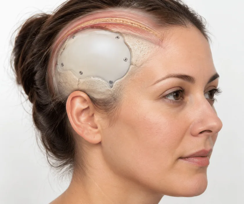
Scalp and skull reconstruction rebuilds the cover of the head after a tumour has been removed. Small defects take a graft or a local flap of scalp. Large ones, or those where bone has been removed, usually need tissue brought from elsewhere in the body with its own blood supply, and sometimes a plate to replace the missing skull.
The scalp has five layers and is held tightly over the skull, which is why a wound that would close easily on the abdomen can be impossible to stitch on the head. Tumours here include skin cancers that have grown deep, tumours arising from the skull itself and cancers that have spread into the bone. Removal often has to take the outer table of bone or the full thickness of it.
Reconstruction has two jobs. The first is stable cover, so that bone, plates or the covering of the brain are never left exposed. The second is a head that looks and feels like a head, with a reasonable outline and a scar that can be hidden by remaining hair where possible.
Choices run from a skin graft on healthy tissue, through rotation flaps that borrow neighbouring scalp, to a free flap taken from the thigh, back or forearm. Where skull has been removed, a shaped plate can restore the contour. Radiotherapy history, defect size and the state of the neck vessels all guide the decision.
This is bigger surgery than most skin cancer work, so fitness and the wider cancer plan matter as much as the defect itself. The decision is usually a joint one with the treating oncologist.
Scans are reviewed with the oncologist and, where bone is involved, with a neurosurgeon. The extent of removal, the likely donor site and the need for a plate are all agreed before the day.
The tumour is removed with a margin, taking bone where it is involved. Frozen section or a later report confirms the edges, and the defect is measured once removal is complete.
A healthy shallow defect may take a graft. A moderate one may take neighbouring scalp rotated across. A deep or irradiated defect usually needs a free flap from the thigh, back or forearm.
For a free flap the artery and vein are joined to vessels in the neck under a microscope. Blood flow is checked repeatedly, because the first day or two are the critical period.
The flap is watched closely in the ward, drains are removed as output falls, and the wider cancer plan such as radiotherapy is restarted once healing allows.
Close monitoring of the flap, drains in place and pain managed with regular medicines. Most people are helped out of bed early, with head position controlled to protect the vessels.
Drains come out, dressings are lighter and stitches are usually removed. Tiredness is common and short walks are encouraged rather than long activity.
The flap has softened and the donor site is comfortable. Further cancer treatment, if it is planned, is often able to start around this stage.
Contour settles, swelling of the flap reduces and any thinning or refinement of the shape can be considered if needed.
Cover is usually reliable, and that is the main measure of success here. Appearance is a compromise, because a flap has no hair and often sits slightly proud of the surrounding scalp before it settles. Many people find that a cap, scarf or hairstyle covers the area comfortably. Contour improves over months, and small refinements are possible once healing is complete.
This is major surgery, often in people who have already had cancer treatment, so the risks deserve a frank discussion before you agree to it.
Recovery continues for weeks after discharge, and the flap needs sensible protection during that period.
Grafts need a healthy bed with blood supply. Over bare bone, a plate or irradiated tissue they usually fail, which is why thicker tissue is brought in instead.
Tissue taken from the thigh or back does not grow scalp hair. Local scalp flaps can carry hair, which is one reason they are preferred when the defect allows.
Modern plates are small and light. Most people carry a card explaining the implant, and daily life is not restricted by it.
Radiotherapy treats the cancer, not the repair. A robust flap is often chosen precisely because it copes better with radiotherapy than thin tissue would.
Elegance Clinic in Surat plans scalp and skull work alongside the treating cancer team, so the repair supports the wider treatment rather than delaying it. Dr. Ashutosh Shah reviews the scans and explains the options and their limits before any commitment.

Free flap reconstruction of the scalp and skull sits in a band because the work is long, needs microsurgery and usually involves a stay of several days. Theatre time, implants such as a shaped plate, intensive monitoring, drains and medicines all contribute.
The band quoted here is a guide. After assessment and review of your scans you receive a written estimate for your own case, and the team will explain what documents your insurer usually needs, since reconstruction after cancer removal is often covered.
These are the questions that come up most often in consultation. If yours is not here, send it on WhatsApp and the team will reply, usually the same day.
Ask your question →Because the work varies a great deal between cases. A long microsurgical procedure, a shaped plate, several days of monitoring and drains all move the figure. After your scans are reviewed you get a written estimate for your own case.
It is major surgery and the vessel joins need close watching in the first days. Serious flap loss is uncommon in experienced hands. Risks such as infection, bleeding and chest problems after a long anaesthetic are discussed openly beforehand.
Expect several days, and longer if a free flap is used, because blood flow is monitored closely at first. Drains are removed as output falls. Discharge happens once the wound is settled and you are moving about comfortably.
Tissue brought from the thigh or back does not grow scalp hair, so a bald patch is expected there. Local scalp flaps can carry hair and are preferred when the defect allows. Many people cover the area with a cap.
Often yes, and previous radiotherapy is one of the main reasons a robust flap is chosen. Irradiated tissue heals slowly, so thin grafts tend to fail there while tissue with its own blood supply usually holds.
The plan is made with your oncologist so that further treatment restarts as soon as healing allows, often around six weeks. A stable, healed wound is what makes radiotherapy possible without breakdown.
Bring your scans, biopsy and operation reports, a list of current medicines and details of any previous radiotherapy. The area is examined, options are explained with their limits and a written estimate follows.
Each technique below has its own page explaining how it works and when it is chosen.
Seeing patients from across the city and beyond: read about the practice on the plastic surgeon in Surat page, or check what a written estimate covers on the costs and insurance page.
