Lymphovenous anastomosis joins a blocked lymph channel directly to a nearby vein, giving trapped fluid a new way out of the limb. Results are best while swelling is still mostly fluid and working lymph vessels remain to be found.
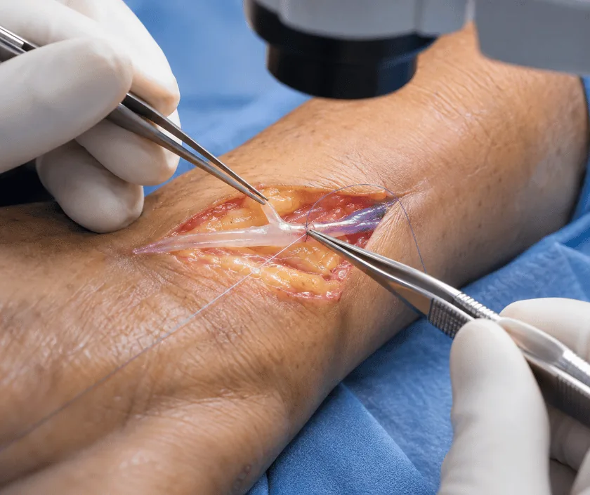
Lymphovenous anastomosis, usually shortened to LVA, is microsurgery in which lymph vessels are stitched into small veins through several short incisions, so lymph drains into the bloodstream instead of pooling. It suits earlier lymphedema where swelling is still fluid predominant. Compression therapy continues afterwards, because the operation improves drainage without restoring a normal lymphatic system.
In a healthy limb, lymph travels through fine vessels up towards nodes in the groin or armpit and then back into the bloodstream. When that route is blocked, vessels below the block stay full while the fluid has nowhere to go. Lymphovenous anastomosis takes advantage of the fact that veins run alongside these lymph vessels and return to the same circulation. Stitch a lymph vessel into a nearby vein, and the fluid bypasses the blockage completely.
Vessels involved are less than a millimetre across, so the work is done under a microscope with instruments and sutures finer than a hair. Before surgery, a dye called indocyanine green is injected under the skin and viewed with a special camera, which shows which lymph channels still carry fluid and where the surgeon should look for them.
Several small incisions are made along the limb, and at each one a suitable lymph vessel and vein are found and joined. Because the incisions are short and no tissue is removed, discomfort is usually modest and the limb keeps its shape. Compression is resumed as the wounds allow, and it stays part of care afterwards.
This is an operation for a particular stage of lymphedema. Selecting the right limb matters far more than surgical enthusiasm, and imaging usually decides the question.
Measurements, staging and imaging come first. Indocyanine green lymphography shows which channels still carry lymph, and the skin is marked where suitable vessels are likely to be found.
Most patients have general anaesthesia, though local anaesthesia with sedation is possible since incisions are small. The limb is positioned so marked sites are easy to reach.
Through incisions usually under three centimetres, the surgeon searches for a lymph vessel and a nearby vein of similar size. Not every site yields a usable pair.
Under the microscope, the lymph vessel is stitched into the vein with extremely fine sutures. Flow is checked, and several joins are made along the limb where possible.
Wounds are closed with fine sutures and dressed. Compression is restarted according to how the wounds are healing, usually within the first days to weeks.
Most patients go home after one night. The limb feels sore around the incisions rather than deeply painful, and it is kept elevated as much as is practical.
Wounds are reviewed and sutures managed. Compression is reintroduced as healing allows, and many patients return to desk work in this period.
Measurements are compared with the baseline. Some limbs have softened noticeably by now, while others change more slowly, and both patterns are seen.
Any real reduction is usually clear by this point. Compression continues, often at a lighter level, and reviews track whether the gain is holding.
Improvement is usually gradual, appearing over months rather than days, and it varies widely between patients. Many find the limb softer and easier to manage with compression, some achieve a clear reduction in size, and a few see little change because the vessels found were not carrying enough flow. Nobody should expect a normal limb or an end to compression therapy after this operation.
Compared with larger operations this is gentle surgery, yet real risks exist and the possibility of limited benefit is the most important of them.
Recovery is straightforward for most patients, and these points protect both the wounds and the new connections.
Almost every patient continues compression afterwards, often at a lighter level. Surgery adds drainage capacity, it does not rebuild the lymphatic system.
Incisions are small but the work is technically demanding, done under a microscope on vessels less than a millimetre wide.
Suitability depends on fluid predominating and channels still carrying lymph. Firm, fatty limbs are better served by operations that remove tissue.
Change is gradual and measured over months. Early weeks mainly involve wound healing rather than visible reduction.
Elegance Clinic in Surat offers lymphovenous anastomosis only where assessment suggests it is the right operation for that limb. Dr. Ashutosh Shah explains what the surgery can add and what will still be required afterwards.

The cost of lymphovenous anastomosis reflects theatre time, microsurgical equipment, the number of sites explored, anaesthesia and the hospital stay. Imaging before surgery and compression garments afterwards are separate items, and both belong in the calculation from the start.
Insurance cover for lymphedema surgery varies by policy and by the underlying cause, which is why cover is assessed case by case. A written estimate is issued before admission, and the team can guide you on the documents insurers usually request.
These are the questions that come up most often in consultation. If yours is not here, send it on WhatsApp and the team will reply, usually the same day.
Ask your question →The band is Rs 1.2L to Rs 2.6L, and where a case falls within it depends on theatre time, how many sites are explored, anaesthesia and length of stay. Imaging beforehand and garments afterwards are separate. A written estimate is issued before admission.
Incisions are small and no tissue is removed, so general risks are lower than for larger operations. Wound infection, bruising and blocked connections remain possible. Fitness for anaesthesia is assessed beforehand, and some patients can avoid a general anaesthetic altogether.
Most patients stay one night and go home with small dressed wounds. Desk work often resumes within about two weeks, while heavier activity waits longer. Compression is restarted as the wounds heal, following the timing you are given.
Improvement is gradual over months and varies considerably. Many patients report a softer limb that responds better to compression, some see clear volume reduction, and a few notice little change. Limbs treated earlier, while still fluid filled, tend to do better.
Yes, in nearly all cases. Surgery adds drainage capacity but does not restore normal lymphatic function. Many patients move to a lighter garment or shorter wearing hours, though stopping altogether usually allows swelling to return.
Once a limb has become firm and fatty, fluid is no longer the main problem and this surgery has little to offer. Active infection, an unhealed wound or continued smoking also postpone or rule it out until those issues are addressed.
Suitability is decided by assessment rather than by symptoms alone. Measurement, staging and lymphatic imaging show whether working channels remain. If they do not, another operation is discussed instead, and the reasoning is explained fully.
Each technique below has its own page explaining how it works and when it is chosen.
Seeing patients from across the city and beyond: read about the practice on the plastic surgeon in Surat page, or check what a written estimate covers on the costs and insurance page.
