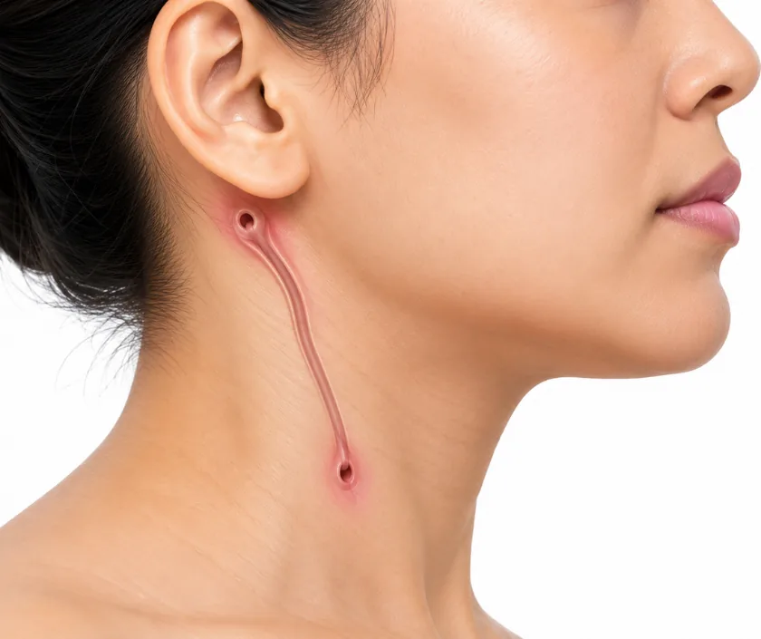
A lump in a child's neck worries most parents, yet many are present from birth and follow a very predictable pattern. During early development the neck folds and fuses in stages. If a small tract or pocket fails to close, it can stay hidden for years and then show itself as a swelling, a tiny opening on the skin, or repeated bouts of infection. Position gives the strongest clue: some lumps sit exactly in the midline, others always appear along the side of the neck.
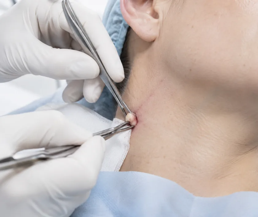
This page explains the main congenital neck lesions and how surgeons tell them apart. Diagnosis at Elegance Clinic in Surat begins with examination, an ultrasound scan and, when needed, further imaging to map how far a tract runs. Removal is usually advised, because these lesions tend to become infected sooner or later and are harder to remove once inflamed. Surgery is planned when the neck is quiet rather than during an active infection.
Location, movement and behaviour separate these lumps far more reliably than size does. That is what guides the scan and the operation chosen.
An ultrasound causes no discomfort, needs no radiation and shows whether a lump is solid, fluid filled or full of channels. It also confirms that normal thyroid tissue is present before a midline cyst is removed, which is an important check in children.
A congenital cyst that becomes infected turns red, tender and hot. Antibiotics settle the episode, but they do not remove the underlying pocket. Surgery is safer and neater once the inflammation has fully calmed, which usually takes several weeks.
Neck skin heals well when incisions follow the natural creases. Scars start pink and firm, then soften over months. Silicone and sun protection help. Children who form thick scars are identified early so the aftercare can be adjusted.
These lesions return when a small piece of tract is left behind. That is why the operation removes more than the visible lump. Previous infection or an earlier incomplete removal makes the tissue planes harder, so the surgeon plans a wider approach.
Most congenital neck lumps can be assessed at a routine appointment. Certain features need faster review.
Short, practical answers to the queries that come up in almost every consultation.
Ask your question →The figure depends on the lesion, the scans needed, the type of anaesthetic and whether the child stays overnight. A cyst removed as a day case costs less than a wide tract dissection. An itemised estimate follows the assessment.
It is a routine procedure in experienced hands, done under general anaesthetic with full monitoring. Risks include bleeding, infection, scar thickening and, rarely, injury to a nearby nerve. Careful mapping beforehand reduces the chance of these problems.
Children usually go home the same day or the next morning. Discomfort settles within a few days with simple pain relief. Normal eating restarts almost immediately, and most return to school within a week, avoiding sport for a little longer.
Incisions are placed in a natural neck crease so the line settles into a shadow over time. Early redness is normal and fades across several months. Sun protection and gentle massage improve the final appearance in most patients.
Not always. Small, quiet preauricular pits and some skin tags can simply be watched. Cysts and tracts are usually removed, because repeated infection makes later surgery harder and leaves more scarring than a planned operation would.
Breathing difficulty, a rapidly enlarging swelling, high fever with redness, or a change in voice needs same day medical care. A lump that has been unchanged for months is not urgent and can wait for a planned appointment.
Bring previous scan reports, any photographs showing the lump at its largest, a list of past infections and current medicines. Those details often change the plan, particularly if there has been an earlier procedure on the same area.