The nose is built from several separate pieces of tissue that swing together and fuse in the middle of the face during early pregnancy. Because so many parts must meet exactly, differences here take many forms: a flattened nostril beside a cleft lip, a lump or pit on the bridge, a passage blocked at the back, or a bridge that never developed height.
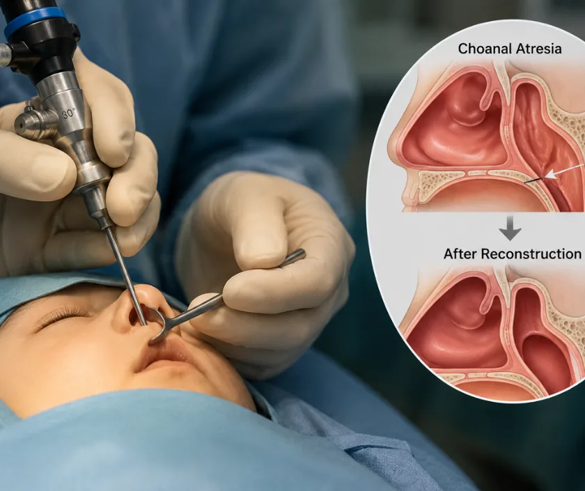
Two things make a child nose different from an adult one. It carries growth centres that keep working through the teenage years, and it is the main route for air in a small baby who has not yet learned to breathe through the mouth. Both facts shape the advice you will be given about timing. This page explains the main groups and their usual handling, and Elegance Clinic in Surat plans nasal work around a child growth rather than against it.
Some of these are questions of appearance that can wait for years, while others affect breathing from the first hours of life. Sorting them apart is the first task at consultation.
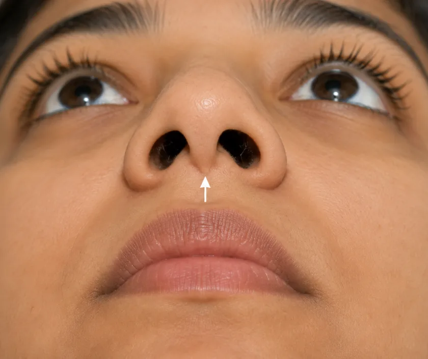
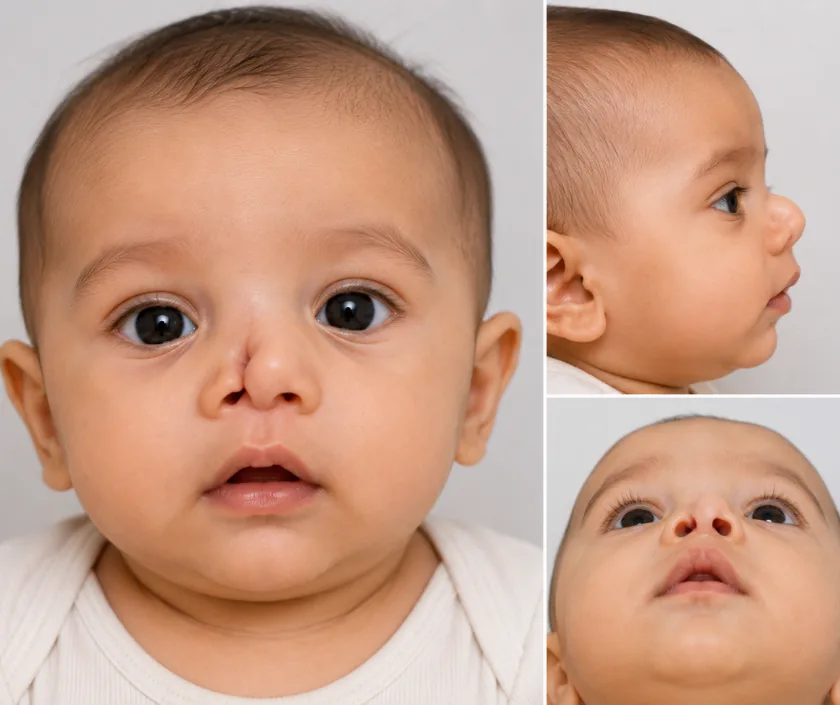
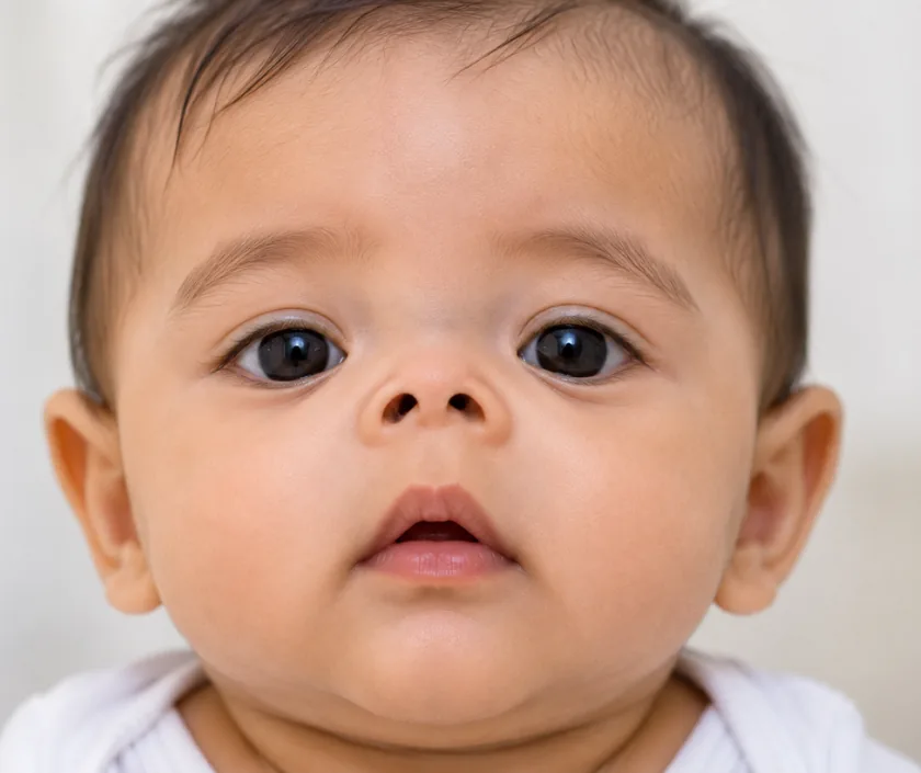

Cartilage in the middle of the nose helps drive facial growth, so heavy surgery there in a young child can hold the midface back. Limited work is done early when breathing demands it. Full reshaping generally waits until the face has finished growing in the late teens.
Parents describe snoring, a blocked sound or mouth breathing, and those observations matter. Examination with a small camera, along with a look at the tonsils and adenoids, shows where the obstruction truly sits. Often the nose is only part of the picture, and treating it alone would disappoint.
A dermoid cyst or similar midline swelling may connect through a bony channel to the lining of the brain. Cutting it out without imaging risks a serious complication. A scan settles the question first, and the operation is then planned with the right team present.
Nasal reconstruction in children is rarely finished in one sitting. Tissue is moved, allowed to settle, then refined. Families are told this at the outset so that a planned second stage feels expected rather than a setback, and each stage is timed around school terms where possible.
Most nasal differences are dealt with on a planned basis. These signs point to an airway or a deeper problem.
These come up at nearly every appointment. Anything specific to your child follows from examination.
Ask your question →It depends heavily on what is being treated. Releasing a blocked passage differs from a staged reconstruction using rib cartilage. Scans, anaesthesia and hospital stay are counted separately. You receive an itemised written estimate once the diagnosis is confirmed and the plan agreed.
Careful, limited surgery is done safely in children when breathing requires it. The concern with major reshaping is that it can restrain growth of the midface, which is precisely why timing is discussed so carefully. Bleeding, infection and the need for revision are the usual risks.
Swelling around the nose settles most noticeably in the first couple of weeks, though the finer shape keeps refining over many months. School usually restarts within a fortnight after minor work. Contact sports, swimming and anything that risks a knock wait considerably longer.
Clear improvement in shape and symmetry is a fair expectation, especially where a cleft has widened one nostril. Exact matching of the two sides is not realistic, since the underlying bone and cartilage differ. Photographs at each stage help everyone judge progress honestly.
Often yes, when the issue is appearance alone. Waiting until growth finishes gives a more stable result and avoids repeated operations. Breathing problems and midline lumps are the exceptions, since both need attention at the age they are found.
A newborn who cannot breathe through the nose needs hospital assessment straight away, because babies depend on nasal breathing during feeds. Sudden swelling, fever or discharge from a midline pit also needs prompt review. A slightly crooked nose in a healthy child does not.
The outside and inside of the nose are examined, sometimes with a small camera, and breathing is assessed on both sides. Old photographs and any previous scans help. You will leave knowing whether the problem is structural, functional or both, and when treatment should be scheduled.
